Fetal Echocardiography in Mumbai, India
Pregnancy is a beautiful yet vulnerable time, and expecting parents often face anxiety when it comes to the baby’s health—especially the heart. Fetal echocardiography is a vital tool that allows doctors to examine a baby’s heart before birth, helping detect congenital heart defects early and guide timely treatment.
Globally, congenital heart disease (CHD) affects 8–10 in every 1,000 live births. In India, it’s estimated that over 2 lakh babies are born annually with some form of CHD. Sadly, many of these conditions remain undiagnosed until it’s too late to intervene effectively. That’s where fetal echocardiography becomes a life-changing advancement.
Dr. Prashant Bobhate, a prominent pediatric cardiologist in Mumbai, shares:
“Fetal echocardiography isn’t just about early diagnosis—it’s about giving families time. Time to prepare, to understand, and to intervene if necessary. The goal is to ensure the baby has the best possible start, even before taking the first breath.”
As Dr. Prashant Bobhate, a leading Pediatric Cardiologist in Mumbai, India, explains:
“Heart conditions in children are not always visible at birth, but early detection can be life-saving. A child’s heart health influences everything—from growth and development to learning and energy levels. Timely diagnosis and treatment ensure a healthier, more active childhood.”
Contact Us
Curious about what conditions can be picked up before birth? Let’s take a look.
Conditions Detected Through Fetal Echocardiography

Fetal echocardiography uses ultrasound waves to create detailed images of your baby’s heart. It helps identify a range of structural and functional heart issues that may require care before or immediately after birth.
Here are some key conditions it can detect:
Septal Defects
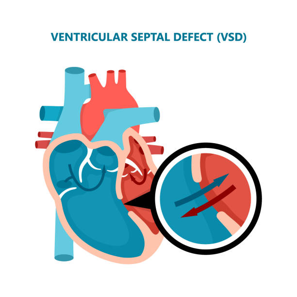
Openings between heart chambers (like ASD or VSD) that can affect blood flow and oxygen delivery.
Hypoplastic Heart Syndrome
Underdevelopment of heart chambers, which needs surgical management shortly after birth.
Transposition of Great Arteries (TGA)
A serious defect where major blood vessels are switched, affecting circulation.
Arrhythmias
Irregular heart rhythms that could indicate an underlying electrical problem.
Valve Malformations
Improperly developed valves that may obstruct or reverse blood flow.

Detecting these early allows neonatal teams to plan interventions, whether it’s delivery at a specialized center or immediate surgery.
Who Should Undergo Fetal Echocardiography?
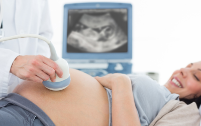
While not routinely advised for all pregnancies, fetal echocardiography is strongly recommended in certain situations where the baby may be at higher risk of heart abnormalities.
It is advised if:
There’s a family history of congenital heart defects or genetic syndromes.
Routine anomaly scans suggest an abnormal heart structure or rhythm.
The mother has pre-existing conditions such as diabetes, lupus, or phenylketonuria.
The mother took certain medications during pregnancy (like anti-seizure drugs or SSRIs).
There are chromosomal anomalies like Trisomy 21 (Down syndrome) seen in other screenings.

In-vitro fertilization (IVF) pregnancies and multiple gestations may also benefit from fetal heart scans.
Abnormal amniotic fluid levels or delayed fetal growth is noted.
Early identification can help families make informed decisions, plan delivery at an equipped center, or prepare emotionally for postnatal care.
Advanced Imaging Techniques Used in Fetal Heart Assessment
While not routinely advised for all pregnancies, fetal echocardiography is strongly recommended in certain situations where the baby may be at higher risk of heart abnormalities.
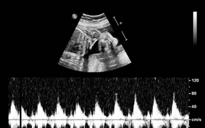
2D Echocardiography
The standard tool for structural heart assessment, showing walls, chambers, and valves in real-time.
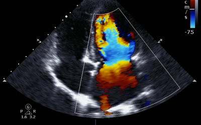
Color Doppler Imaging
Helps assess blood flow direction and speed—crucial for spotting abnormal patterns.
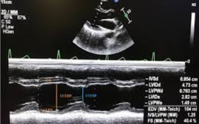
M-Mode Echo
Evaluates fetal heart rhythm and function by measuring wall movement over time.

3D/4D Imaging (when needed)
Offers a more comprehensive view in complex cases, though not always required.
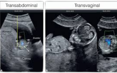
Fetal Transabdominal and Transvaginal Approaches
Depending on gestational age and fetal position, the cardiologist selects the most effective method to get clear images.
Accuracy depends not just on the machine, but on the operator’s training and interpretation. Dr. Bobhate brings years of hands-on expertise in interpreting these scans accurately and compassionately. His expertise makes him a highly sought-after pediatric heart specialist for Fetal echocardiogram in Mumbai.
Dr. Prashant Bobhate Explains the Fetal Echocardiography Process
Dr. Prashant Bobhate, an accomplished fetal echocardiography specialist in Mumbai, describes the process as “non-invasive, informative, and reassuring—when done by the right hands.”
What to expect:
Timing
Best done between 18 to 24 weeks of gestation for optimal clarity and detail.
Duration
The scan takes about 30 to 45 minutes, depending on fetal position and complexity.
Preparation
No fasting or special preparation is needed. It’s just like a regular ultrasound, but more focused.
Procedure
The mother lies on an exam table while the specialist uses a transducer to glide over the belly with gel to capture images.
Results Discussion
Parents are guided through what’s visible, and a report is shared with the referring obstetrician or fetal medicine specialist.
“The test itself is painless, but its value in preparing for life-saving decisions is immeasurable. Whether it confirms normalcy or highlights a concern, it gives families the clarity they need.”
Why Choose Dr. Prashant Bobhate for Fetal Echocardiography in Mumbai?
Dr. Prashant Bobhate is recognized as one of the top fetal echocardiogram experts in Mumbai—for good reason. His reputation rests on both clinical precision and emotional sensitivity.
Dual Expertise in Pediatrics & Cardiology:
Deep understanding of both fetal and postnatal heart function ensures continuity of care.
Specialized Training:
Trained at prestigious national centers, including Escorts Heart Institute.
Over 12 Years of Focused Experience:
Having managed complex cases in infants and fetuses, he brings rare insight to every scan.
Founder of a Pulmonary Hypertension Clinic:
His work in early detection reflects his long-term commitment to preventive cardiology.
Advanced Technology Access:
Performs scans with the latest echo machines at Kokilaben Hospital—ensuring clarity and safety.
Collaborative Approach:
Works closely with obstetricians, neonatologists, and fetal medicine experts for integrated care.
Parental Counseling:
Known for his clear, honest communication—empowering families with knowledge, not fear.
Award-Winning Excellence:
Honored by national and international cardiology societies for his research and presentations.
Still have questions about fetal echo? We’ve got you covered.
FAQs
Which week is best for fetal echocardiography?
The ideal window is between 18 to 24 weeks of pregnancy. This period allows the baby’s heart to be developed enough for detailed assessment while still offering time for planning, if needed.
What is the cost of fetal echo?
The fetal echo cost in Mumbai typically ranges between ₹3,500 to ₹7,000, depending on the facility and the expertise involved. It’s advisable to choose centers that have specialized pediatric cardiologists for optimal accuracy.
Does every baby get a fetal echo?
No, it’s not a routine scan. Fetal echocardiograms are recommended when risk factors are present—either maternal, familial, or based on prior ultrasound findings.
Why do doctors recommend fetal echo?
Doctors recommend it to:
- Detect structural heart defects
- Evaluate abnormal rhythms
- Plan neonatal interventions
- Support timely medical or surgical planning
- Offer reassurance in high-risk pregnancies
Can fetal echo detect Down syndrome?
Not directly. However, certain heart defects, such as atrioventricular septal defects, are more common in babies with Down syndrome. If these are spotted, further genetic testing may be advised.
What 5 abnormalities can be found on the echocardiogram?
Common abnormalities include:
- Atrial Septal Defect (ASD)
- Ventricular Septal Defect (VSD)
- Hypoplastic Left Heart Syndrome (HLHS)
- Transposition of the Great Arteries (TGA)
- Tetralogy of Fallot (TOF)
Each condition varies in severity, but early diagnosis improves planning and treatment outcomes significantly.
Disclaimer: The information shared in this content is for educational purposes only and not for promotional use.

