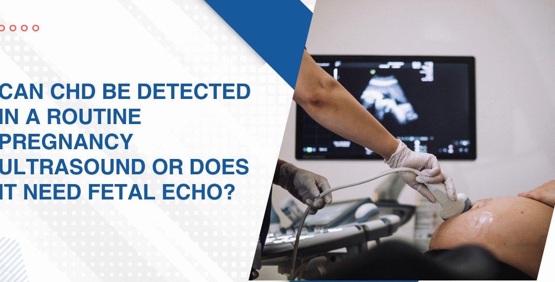
Can CHD Be Detected in a Routine Pregnancy Ultrasound or Does It Need Fetal Echo?

Can CHD Be Detected in a Routine Pregnancy Ultrasound or Does It Need Fetal Echo?

Congenital heart defects can often be suspected during a routine mid-trimester ultrasound between 18 and 22 weeks which includes a four-chamber heart view. However a specialised fetal echocardiogram is required for definitive detailed diagnosis because it is significantly more sensitive and specific for detecting complex cardiac abnormalities that the standard anomaly scan simply wasn’t designed to find.

“A normal anomaly scan is reassuring but it is not a cardiac clearance and families who come to me after a normal 20-week scan and a baby born with a significant defect are not rare. The anomaly scan looks at the heart. Fetal echo looks into it,” says Dr. Prashant Bobhate, Pediatric Cardiologist in Mumbai, India.

What Can a Routine Pregnancy Ultrasound Actually Detect?

The anomaly scan was designed to screen the whole fetus across multiple organ systems in a single appointment and cardiac assessment is one part of that broader sweep not the focus of it.

  • Four-chamber view: The standard anomaly scan includes a four-chamber view that can identify major abnormalities in chamber size and symmetry but this single view misses outflow tract defects, valve abnormalities and many conotruncal anomalies that only become visible with additional dedicated cardiac views.
  • Major structural defects: Large AVSDs, significant hypoplastic left heart and major ventricular asymmetry are visible on a well-performed anomaly scan in experienced hands but detection rates drop significantly in less specialised settings where cardiac views are obtained quickly as part of a broader structural survey.
  • Operator dependence: The quality of cardiac screening on a routine ultrasound depends entirely on the operator’s training, the equipment available and the fetal position on the day and the same defect can be missed on one scan and flagged on another depending entirely on those variables.
  • What it cannot show: Outflow tract abnormalities including TOF, transposition and pulmonary atresia, most valve defects, abnormal venous connections and many rhythm abnormalities simply cannot be reliably assessed on a routine anomaly scan regardless of how carefully it is performed.

Every pregnancy with a risk factor for congenital heart disease needs a dedicated fetal echocardiography assessment that goes well beyond what the anomaly scan was designed to provide.

When Is a Fetal Echo Specifically Needed?

Not just for high-risk pregnancies, fetal echocardiography is important for any pregnancy where the anomaly scan raises a concern

  • Abnormal anomaly scan: Any cardiac finding flagged on routine ultrasound including an asymmetric four-chamber view, an unusual cardiac axis or a suspected outflow tract abnormality is an immediate indication for fetal echo by a specialist who does this regularly not occasionally.
  • Family history of CHD: A parent or sibling with a congenital heart defect increases the recurrence risk to 3 to 5 percent and that risk is high enough to warrant fetal echo in every pregnancy in that family regardless of what the routine scan shows.
  • Maternal conditions: Diabetes, systemic lupus, phenylketonuria and certain autoimmune conditions are associated with specific fetal cardiac abnormalities and mothers with these conditions need fetal echo as a standard part of antenatal care not an optional add-on decided case by case.
  • Fetal arrhythmia on Doppler: A sustained irregular fetal heart rhythm picked up on routine Doppler needs fetal echo to characterise whether it’s a benign ectopic beat pattern or a sustained arrhythmia that requires monitoring or treatment before delivery.

Parents wanting to understand what fetal echo can and cannot detect and how findings change management before birth should read this piece on can a fetal echo detect baby heart defects because understanding the scope of the investigation is what allows families to ask the right questions at the right appointment.

Why Choose Dr. Prashant Bobhate for PH Management in Mumbai?

A fetal echo is only as useful as the person reading it and a specialist who performs fetal echos occasionally as part of a general obstetric practice reads the images very differently from one who has spent years specifically in fetal and paediatric cardiology managing the full spectrum of what those images reveal before birth and after. Dr. Prashant Bobhate has spent over 12 years performing fetal echocardiography and managing the post-natal cardiac outcomes of defects detected before birth at the Children’s Heart Centre, Kokilaben Dhirubhai Ambani Hospital. Escorts Heart Institute New Delhi.

Schedule a consultation to find out if a cure is possible and what the right treatment plan looks like for you.

FAQs

At what week should a fetal echo be done?

Between 18 and 24 weeks is the optimal window for most structural assessments though an experienced centre can obtain useful information from 16 weeks onwards and earlier scans may be indicated in specific high-risk situations.

Is fetal echo safe for the baby?

Yes completely because it uses the same ultrasound technology as a routine pregnancy scan with no radiation involved and no known risk to the fetus at the frequencies and durations used in clinical fetal cardiac assessment.

Can a fetal echo miss heart defects?

Some defects including small VSDs, mild valve abnormalities and some arrhythmias can be missed or only become apparent after birth which is why newborn cardiac assessment remains important even after a normal fetal echo.


Does a normal anomaly scan mean the baby's heart is definitely fine?

No because the anomaly scan is a structural survey not a cardiac assessment and a normal four-chamber view at 20 weeks does not exclude outflow tract defects, valve abnormalities or arrhythmias that fetal echo specifically looks for.

References:

What Causes Sudden Cardiac Death In Young Athletes?

What Causes Sudden Cardiac Death In Young Athletes?

A diet for children with pulmonary hypertension should focus on low-sodium, nutrient-dense whole foods that support heart and lung function. Key priorities include limiting processed food and added salt, ensuring adequate protein for muscle strength, managing total fluid intake and maximising vitamins C, D and iron.

“Families focus almost entirely on medications and rightly so but nutrition is the one thing happening three times a day every day that nobody is optimising and in a child with pulmonary hypertension that’s a significant missed opportunity for supporting the heart,” says Dr. Prashant Bobhate, Pediatric Cardiologist in Mumbai, India.

What Are the Most Common Causes of Sudden Cardiac Death in Young Athletes?

There’s a recognisable list and understanding it is what makes pre-participation cardiac screening a clinical necessity rather than an optional extra for competitive sport.

  • Hypertrophic cardiomyopathy: The single most common cause in young athletes where abnormally thickened myocardial fibres create an unstable electrical substrate that generates ventricular fibrillation under the precise haemodynamic stress that intense physical exertion produces.
  • Anomalous coronary arteries: A coronary artery arising from the wrong sinus can course between the aorta and pulmonary artery in a way that compresses the vessel during exercise-induced aortic expansion and cuts off blood supply to a large myocardial territory at exactly the moment demand is highest.
  • Long QT syndrome: The ECG looks almost normal at rest but the prolonged repolarisation interval becomes dangerous under adrenergic stimulation from exercise or sudden emotional stress and can trigger torsades de pointes that degenerates into ventricular fibrillation without any warning symptom.
  • Myocarditis: Viral inflammation of the heart muscle creates zones of electrical instability that persist well beyond the acute illness phase and an athlete who returns to training too soon after a viral illness with unrecognised myocarditis is training on a heart that can no longer reliably maintain normal rhythm under load.

Every young athlete with a family history of sudden cardiac death, unexplained syncope during exercise or a newly detected murmur deserves a formal assessment and pediatric arrhythmia evaluation maps the electrical and structural risk before any return to competitive sport gets cleared.

Can Sudden Cardiac Death in Young Athletes Be Prevented?

Often yes. When the right screening happens before the right sport at the right intensity.

  • Pre-participation ECG: A resting 12-lead ECG picks up Long QT syndrome, WPW pattern, Brugada pattern and HCM-related repolarisation changes that a physical examination alone completely misses and costs a fraction of what treating a survivor of cardiac arrest costs in every dimension imaginable.
  • Echocardiography for high-risk athletes: Any athlete with an abnormal ECG, a family history of sudden cardiac death under 50, unexplained exertional syncope or a cardiac murmur needs an echo before competing at any level where sustained high-intensity effort is involved.
  • Restricting sport in diagnosed conditions: A child diagnosed with HCM, Long QT or anomalous coronary arteries needs a formal sport eligibility assessment because the specific activity restriction required is condition-specific and a blanket ban applied without assessment is as clinically unhelpful as no restriction at all.
  • AED availability at sports venues: Automated external defibrillators at schools, sports academies and training grounds don’t prevent the arrhythmia but they convert a potentially fatal event into a survivable one when used within the first three to five minutes and their absence at Indian sports venues is a preventable gap that costs young lives every year.

Parents wanting to understand what cardiac warning signs in young children look like before any sport-related event forces the issue should read this piece on how to spot the early signs of heart disease in neonates because the cardiac conditions that cause sudden death in athletes at sixteen were present at birth and detectable long before the first sprint.

Why Choose Dr. Prashant Bobhate for PH Management in Mumbai?

Pre-participation cardiac screening for a young athlete isn’t a routine check. It’s reading an ECG in the context of the sport being played, the intensity involved and the specific conditions that that particular child’s family history or symptom profile makes worth looking for. Not a form-filling exercise. A real assessment. Dr. Prashant Bobhate has spent over 12 years managing paediatric arrhythmia, hypertrophic cardiomyopathy, sudden cardiac death risk stratification and sport eligibility assessments across every age group at the Children’s Heart Centre, Kokilaben Dhirubhai Ambani Hospital. Escorts Heart Institute New Delhi.

Schedule a consultation to find out if a cure is possible and what the right treatment plan looks like for you.

FAQs

How common is sudden cardiac death in young athletes in India?

Exact figures are underreported because many events are attributed to heat stroke or exhaustion without cardiac investigation but HCM and arrhythmia syndromes are consistently identified as the leading causes wherever proper post-event evaluation is carried out.

Should every child have a cardiac screening before playing competitive sport?

Yes at minimum an ECG and clinical history because the conditions that cause sudden cardiac death in athletes are detectable before the event and a normal screening is a genuinely reassuring baseline that parents and coaches both deserve to have.

Can a child with a heart condition ever play sport?

Many can with the right assessment and specific guidance because not every cardiac condition carries the same exercise risk and a formal sport eligibility assessment gives a child and family a real answer rather than a blanket restriction based on diagnosis alone.


What should parents do if their child faints during sport?

Treat it as a cardiac event until proven otherwise because exertional syncope in a young athlete is always a red flag that needs same day cardiac assessment including ECG and echo before the child returns to any physical activity.

References:

Diet and Nutrition For Children With Pulmonary Hypertension

Diet and Nutrition For Children With Pulmonary Hypertension

A diet for children with pulmonary hypertension should focus on low-sodium, nutrient-dense whole foods that support heart and lung function. Key priorities include limiting processed food and added salt, ensuring adequate protein for muscle strength, managing total fluid intake and maximising vitamins C, D and iron.

“Families focus almost entirely on medications and rightly so but nutrition is the one thing happening three times a day every day that nobody is optimising and in a child with pulmonary hypertension that’s a significant missed opportunity for supporting the heart,” says Dr. Prashant Bobhate, Pediatric Cardiologist in Mumbai, India.

What Should Children With Pulmonary Hypertension Be Eating?

The goal is calorie-dense, nutrient-rich food that doesn’t overload the circulation, supports cardiac muscle function and maintains a body weight the right ventricle can actually sustain without additional strain.

  • Adequate calories: Children with PH burn more calories than healthy peers because increased work of breathing and cardiac effort raises resting energy expenditure and underfeeding a child with PH isn’t cautious it’s actively harmful to the right ventricle’s ability to cope.
  • Protein for muscle strength: Adequate dietary protein from eggs, lentils, dairy and lean meats supports respiratory muscle strength and prevents the muscle wasting that significantly worsens functional capacity and exercise tolerance in children with chronic cardiopulmonary disease.
  • Vitamins C and D: Vitamin C from fresh fruits and vegetables supports vascular integrity and immune function while vitamin D deficiency is common in children with chronic cardiac disease and contributes to muscle weakness, fatigue and poor bone health that compounds the disease burden.
  • Iron-rich foods: Many children with cyanotic PH develop iron deficiency alongside erythrocytosis and ensuring adequate dietary iron through lentils, green leafy vegetables and fortified cereals supports healthy red cell production without the risks of unmonitored supplementation.

Understanding how nutrition fits into the broader management picture for children with pulmonary hypertension is exactly what a thorough medical management of pulmonary hypertension consultation covers alongside medication planning and follow up.

What Should Children With PH Avoid in Their Diet?

Specific things make the right heart’s job harder. Worth knowing clearly.

  • High sodium foods: Processed snacks, packaged noodles, pickles, papads and restaurant food carry sodium loads that drive fluid retention in children whose kidneys are already responding to reduced cardiac output by retaining more salt and water than they should.
  • Large meals: Big meals shift significant blood volume to the gut during digestion and in children with limited cardiac reserve that sudden redistribution triggers breathlessness, fatigue and reduced oxygen saturation so smaller more frequent meals across the day are always the better approach.
  • Grapefruit: Grapefruit and grapefruit juice inhibit the enzyme that metabolises several PH medications including sildenafil and bosentan and consuming it regularly alters effective drug levels in ways that are genuinely unpredictable and clinically significant for a child already on a carefully titrated regimen.
  • Caffeine: Caffeinated drinks including cola, energy drinks and strong tea can trigger arrhythmias and raise heart rate in children whose cardiac rhythm is already more vulnerable than in a healthy child and these simply don’t belong in a PH child’s regular diet.

Parents wanting to understand the broader treatment approach for children on active PH therapy should also read about PH differences, as understanding related heart and lung conditions helps families better follow long-term management decisions

Why Choose Dr. Prashant Bobhate for PH Management in Mumbai?

Managing paediatric pulmonary hypertension requires more than medication alone. With over 12 years of experience, Dr. Prashant Bobhate provides comprehensive PH care including nutrition, growth monitoring, activity guidance, and long-term home management support at leading cardiac centres including Kokilaben Dhirubhai Ambani Hospital and Escorts Heart Institute New Delhi.

Schedule a consultation to find out if a cure is possible and what the right treatment plan looks like for you.

FAQs

Should children with pulmonary hypertension follow a low-salt diet?

Yes because excess sodium drives fluid retention that increases circulating blood volume and adds pressure on the right heart and reducing processed food, added salt and high-sodium snacks is one of the most practical daily interventions families can make.

Can children with PH eat normally at school?

Mostly yes with specific adjustments around sodium, large meal sizes and caffeinated drinks and the school canteen and tiffin need to reflect those adjustments rather than treating school meals as separate from the management plan.

Is grapefruit actually a problem for children on PH medication?

Yes, because grapefruit inhibits the enzyme that breaks down several PH medications including sildenafil and bosentan and consuming it regularly alters effective drug levels in ways that are unpredictable and clinically significant.

Do children with pulmonary hypertension need more calories than healthy children?

Usually, yes because the increased work of breathing and cardiac effort raises resting energy expenditure and underfeeding a child with PH actively worsens right heart function rather than protecting it.

References:

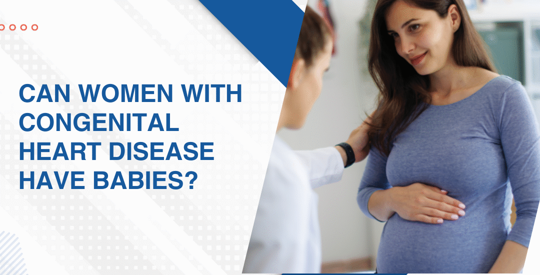
Can Women With Congenital Heart Disease Have Babies?

Can Women With Congenital Heart Disease Have Babies?

Yes, Most women with congenital heart disease can safely have children but pregnancy carries real risks including arrhythmia, heart failure and stroke that vary significantly by defect type and repair history. It requires specialised care from a team including a cardiologist and maternal-fetal medicine specialist and that conversation needs to start before conception not after a positive test arrives.

“I’ve seen women with complex congenital heart disease carry pregnancies to term and do well and I’ve seen women with seemingly simple repaired defects run into problems nobody anticipated because nobody did a proper pre-conception cardiac evaluation before they conceived,” says Dr. Prashant Bobhate, Pediatric Cardiologist in Mumbai, India.

What Makes Pregnancy Risky for Women With Congenital Heart Disease?

Pregnancy puts the cardiovascular system under a sustained haemodynamic load and a heart already managing abnormal anatomy handles that load very differently from a structurally normal one.

  • Blood volume surge: Pregnancy increases blood volume by 40 to 50 percent and a heart with limited functional reserve can decompensate rapidly under that sustained demand even if it was compensating adequately before conception.
  • Arrhythmia risk: The haemodynamic and hormonal changes of pregnancy lower the arrhythmia threshold and atrial flutter, SVT and ventricular arrhythmias can emerge in women who had no rhythm history whatsoever before conceiving.
  • Pulmonary hypertension: Women with pulmonary arterial hypertension face the highest maternal mortality risk of any cardiac condition in pregnancy because systemic vasodilation against fixed pulmonary vascular resistance can deteriorate rapidly and catastrophically.
  • Fontan circulation: The passive pulmonary blood flow that sustains a Fontan depends on venous pressure gradients that pregnancy disrupts and obstetric outcomes in Fontan patients need the most experienced multidisciplinary team available.

Every woman with a congenital heart diagnosis considering pregnancy deserves a full structural and functional assessment and congenital heart disease evaluation before conception maps current anatomy, ventricular function and pulmonary pressures before any pregnancy planning moves forward.

What Does Safe Pregnancy Look Like for Women With CHD?

Planned, monitored and never managed alone by a single specialist.

  • Pre-conception counselling: A cardiac assessment before attempting pregnancy establishes the current baseline of ventricular function, pulmonary pressure and rhythm status so any changes during pregnancy can be measured against something real rather than guessed at.
  • Medication review: Several cardiac medications including warfarin, ACE inhibitors and some antiarrhythmics carry real fetal risk and switching to pregnancy-safe alternatives before conception is far safer than scrambling to change them after a positive test.
  • High-risk obstetric co-management: A maternal-fetal medicine specialist needs to be part of the team from the start because delivery timing, mode of delivery and anaesthetic approach all have cardiac implications that require both teams aligned well before the moment arrives.
  • Delivery planning: Women with significant CHD need delivery at a centre with cardiac anaesthesia, adult congenital cardiology and a cardiac ICU on site because the haemodynamic shift immediately after delivery is one of the highest-risk moments in the entire pregnancy.

Parents wanting to understand the long-term journey of congenital heart care should also explore surgery costs, as treatment planning often begins early and financial preparation plays an important role in managing cardiac care from childhood onward

Why Choose Dr. Prashant Bobhate for Cardiac Care in Pregnancy With CHD in Mumbai?

A woman with congenital heart disease who wants to have children needs a cardiologist who understands what her specific anatomy does under the haemodynamic load of pregnancy, can advise on medication changes before conception and can coordinate with the obstetric team across the full nine months rather than showing up only when something goes wrong. Dr. Prashant Bobhate has spent over 12 years managing adult congenital heart disease including pre-conception counselling and pregnancy planning for women with complex repaired and unrepaired cardiac conditions at the Children’s Heart Centre, Kokilaben Dhirubhai Ambani Hospital.

Schedule a consultation to find out if a cure is possible and what the right treatment plan looks like for you.

FAQs

Is it safe to get pregnant with a repaired congenital heart defect?

For many women yes but safety depends on the specific defect, repair type, current ventricular function and pulmonary pressures and needs a formal assessment not an assumption based on how well the woman feels.

Which congenital heart conditions make pregnancy most dangerous?

Pulmonary arterial hypertension carries the highest maternal mortality risk followed by unrepaired cyanotic defects, Fontan circulation and severe left heart obstruction which all need the most intensive pre-pregnancy evaluation.

Can a woman with congenital heart disease pass it to her baby?

Yes with most congenital cardiac conditions carrying a 3 to 5 percent recurrence risk in offspring and fetal echocardiography during pregnancy is recommended for all women with a known congenital heart diagnosis.

When should a woman with CHD start planning for pregnancy?

At least six to twelve months before attempting conception to allow time for cardiac assessment, medication review and specialist team coordination before any haemodynamic stress begins.

References:

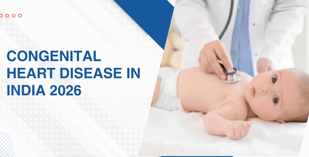
Congenital Heart Disease In India 2026

Congenital Heart Disease In India 2026

Around 240,000 babies are born with congenital heart disease in India every year, roughly one in every 100 newborns, making it the leading cause of birth defect-related deaths and accounting for nearly 10 percent of all infant deaths in the country. The defects are present. The surgical solutions exist. The gap that kills children sits between those two facts and in 2026 that gap is still far wider than it should be.

“The numbers are not abstract to me. Every year tens of thousands of Indian children who could have been saved with a timely intervention aren’t and the reason is almost never that the surgery was too difficult. It’s that nobody found them early enough or the family couldn’t access the right centre in time” says Dr. Prashant Bobhate, Pediatric Cardiologist in Mumbai, India.

What Do the Numbers Actually Look Like in India in 2026?

It creates a deliberate right-to-left shunt at the level of the great vessels that allows the overloaded right ventricle to decompress directly into the aorta rather than continuing to push against a pulmonary vascular resistance it can no longer overcome.

  • Right ventricular decompression: The shunt allows blood to bypass the high-resistance pulmonary circulation entirely which immediately reduces the pressure load on a right ventricle that was heading toward failure and gives the chamber a chance to recover function it was rapidly losing.
  • Maintains cardiac output: A failing right ventricle in severe PH can no longer maintain adequate systemic circulation and the Potts shunt preserves overall cardiac output even at the cost of some arterial oxygen desaturation which is an acceptable trade-off when the alternative is right heart collapse.
  • Bridge to transplant: In children who are maxed out on medical therapy and deteriorating despite everything, the Potts shunt buys enough haemodynamic stability to reach lung transplant listing criteria and survive the wait in a condition where transplant is actually still feasible.
  • Transcatheter approach: India’s first successful transcatheter Potts shunt was performed at this centre using a catheter-based technique rather than open surgery which reduces the procedural risk significantly in children who are already haemodynamically fragile and couldn’t safely tolerate a sternotomy at that point in their disease.

Understanding what interventional options exist for children with severe pulmonary hypertension who have exhausted conventional medical pathways is exactly what a thorough interventional treatment for pulmonary hypertension assessment maps out before any decision gets made.

Which Children With PH Actually Need a Potts Shunt?

Better than a decade ago, but still nowhere near where they should be

  • Better Surgical Outcomes: Leading Indian paediatric cardiac centres now achieve outcomes comparable to global standards for common congenital heart surgeries when treated early
  • Delayed Diagnosis Risk: Many children reach surgery late with complications like malnutrition or pulmonary hypertension, which worsens recovery
  • Ayushman Bharat Support: PM-JAY has improved affordability, but specialist care remains concentrated in limited cities
  • Fetal Diagnosis Advantage: Early detection through fetal echocardiography improves outcomes by enabling timely referral before complications develop 

Parents wanting to understand why fetal cardiac detection matters should read about the importance of fetal diagnosis of critical congenital heart disease, as early detection can mean the difference between planned care and an emergency after birth.

Why Choose Dr. Prashant Bobhate for Potts Shunt and Severe PH Care in Mumbai?

The statistics describe a system problem. What matters to any individual family is whether the specialist in front of them has the training, the equipment and the experience to find the defect early, make the right call about timing and intervention and follow that child for as long as the heart needs watching. Dr. Prashant Bobhate has spent over 12 years managing congenital heart disease across every defect type and every level of complexity at the Children’s Heart Centre, Kokilaben Dhirubhai Ambani Hospital, one of Mumbai’s leading paediatric cardiac centres with fetal echo, interventional cardiology and surgical co-management all in one place

Schedule a consultation to find out if a cure is possible and what the right treatment plan looks like for you.

FAQs

Is the Potts shunt a cure for pulmonary hypertension in children?

Around 240,000 babies are born with congenital heart disease in India annually and of these roughly 50,000 have defects serious enough to need intervention within the first year of life.

What is the survival rate for children with congenital heart disease in India?

At high-volume specialist centres outcomes for standard repairs are now comparable to international benchmarks but delayed diagnosis, malnutrition at presentation and limited specialist access still significantly worsen outcomes for children outside major urban centres.

Which is the most common congenital heart defect in Indian children?

Ventricular septal defect is the most commonly diagnosed congenital heart defect in Indian children followed by atrial septal defect and these acyanotic lesions account for the large majority of CHD cases seen at tertiary cardiac centres.

Does Ayushman Bharat cover congenital heart surgery for children in India?

Yes for several procedures at empanelled hospitals but geographic concentration of surgical expertise and gaps between scheme package rates and actual procedure costs at private centres mean coverage on paper doesn’t always translate into accessible care in practice.

References:

Call Now Button