What Is Rheumatic Heart Disease And How Is It Caused n Children

What Is Rheumatic Heart Disease And How Is It Caused n Children

Rheumatic heart disease is permanent valve damage caused by rheumatic fever, itself triggered by an untreated group A streptococcal throat infection. The immune system meant to fight the bacteria turns on the heart valves instead and that misdirected attack leaves scar tissue that stiffens and narrows valves in ways the heart simply can’t recover from without intervention.

“The tragedy of rheumatic heart disease is that it’s almost entirely preventable and yet I still see children with severely damaged mitral valves from a strep throat written off as a viral fever three or four years earlier,” says Dr. Prashant Bobhate, Pediatric Cardiologist in Mumbai, India.

What Actually Causes Rheumatic Heart Disease in Children?

The bacteria infects the throat and if nobody treats it properly the immune response that follows doesn’t stay where it belongs.

  • Strep ignored: An untreated streptococcal throat infection triggers antibodies that cross-react with cardiac tissue because the bacterial proteins look enough like heart valve proteins to confuse the immune system completely.
  • Repeated infections: One episode damages the valve somewhat but each recurrent strep infection in a child without penicillin prophylaxis adds another layer of scarring that compounds quietly with every round.
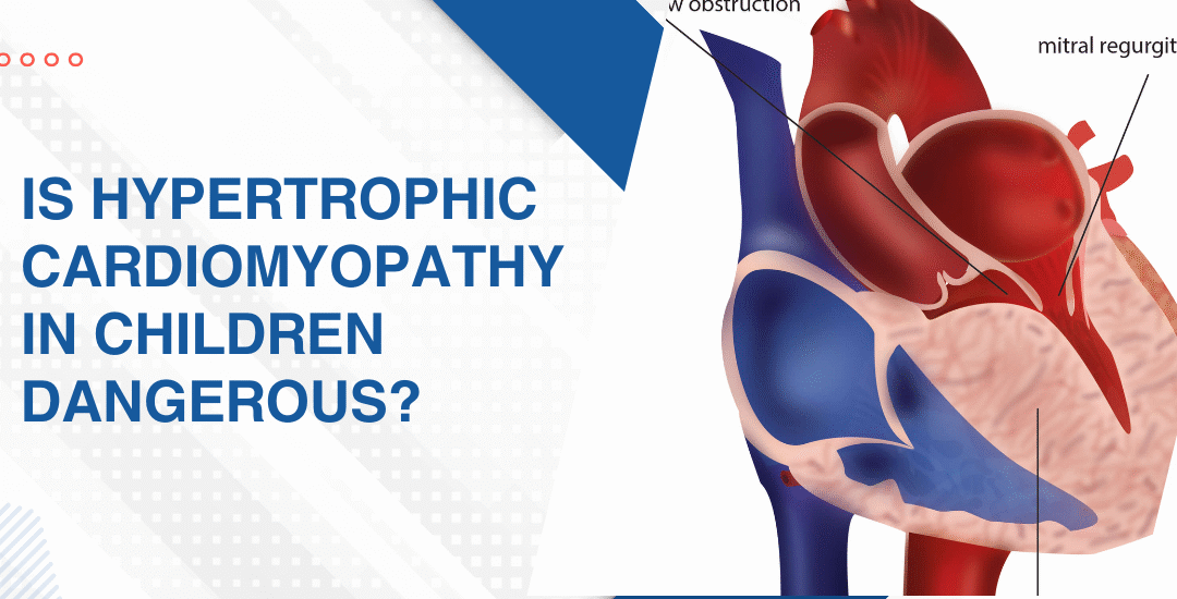
  • Mitral valve: This is the valve that takes the worst hit and the leaflets gradually thicken, fuse at the tips and lose the flexibility they need to open and close across a lifetime of beats.
  • Poverty drives it: Overcrowded homes, no antibiotics nearby and delayed care all mean strep throat goes untreated long enough to start the whole cascade and India carries a disproportionately high burden of this disease for exactly that reason.

Children showing breathlessness, reduced stamina or a murmur after a history of recurrent throat infections need urgent specialist assessment and pediatric heart failure management starts with knowing exactly what the valves look like right now.

How Is Rheumatic Heart Disease Managed in Children?

Prevention first. Then damage control. Surgery only when the valve has gone too far.

  • Penicillin injections: Any child who has had confirmed rheumatic fever needs monthly benzathine penicillin for years, sometimes decades, to stop recurrent strep from adding more damage to what’s already there.
  • Echo surveillance: Regular echocardiography tracks how the valve is changing and catches the point where waiting becomes genuinely riskier than stepping in and doing something about it.
  • Symptom medications: Diuretics and vasodilators manage the haemodynamic load in symptomatic children who aren’t yet at the threshold where a procedure becomes the better option.
  • Balloon or surgery: When mitral stenosis is severe enough to impair output and daily life, balloon dilatation or surgical repair is the only real way to restore forward flow and give the left atrium some relief.

Parents wanting to understand what cardiac warning signs look like in children before any formal valve diagnosis gets made should read this piece on top 5 warning signs of pediatric heart failure because catching it early is always what keeps the most options open.

Why Choose Dr. Prashant Bobhate for Rheumatic Heart Disease Care in Mumbai?

You want someone who can look at the valve on echo, tell you honestly where it’s heading and have the real conversation about what twelve looks like now and what twenty-five looks like if nothing changes. Not a soft answer designed to make the appointment feel better. A plan. Dr. Prashant Bobhate has spent over 12 years managing paediatric valve disease from early subclinical rheumatic damage through severe mitral involvement requiring intervention at the Children’s Heart Centre, Kokilaben Dhirubhai Ambani Hospital.

Schedule a consultation to find out if a cure is possible and what the right treatment plan looks like for you.

FAQs

Can rheumatic heart disease be cured in children?

The damage already done can’t be undone but progression stops with consistent penicillin prophylaxis and severely damaged valves can be repaired or replaced when intervention becomes necessary.

How common is rheumatic heart disease in India?

Very common. Delayed antibiotic access, overcrowding and late diagnosis mean strep throat progresses to rheumatic fever here far more often than it should and India carries one of the highest RHD burdens globally.

What are the first signs of rheumatic heart disease in a child?

Breathlessness during activity, reduced stamina, joint pains after a recent throat infection or a newly detected murmur are the signs that should send a family straight to a paediatric cardiologist without waiting.

Does every child with rheumatic fever develop heart disease?

Not every child but around 30 to 45 percent with untreated rheumatic fever develop some valve damage which is exactly why penicillin prophylaxis after any confirmed episode isn’t optional.

References:

Is Hypertrophic Cardiomyopathy In Children Dangerous?

Is Hypertrophic Cardiomyopathy In Children Dangerous?

Yes. Hypertrophic cardiomyopathy is a leading cause of sudden cardiac death in young athletes and children, making it one of the most serious cardiac diagnoses a family can receive. But dangerous doesn’t mean hopeless. Most children with a confirmed HCM diagnosis can lead normal lives with proper treatment, careful monitoring and a management plan built around their specific risk profile by a paediatric cardiologist who actually understands the condition.

“HCM is one of those diagnoses that families come to me genuinely not knowing what to do with because the child looks well, plays normally and then you show them the echo and they can’t reconcile the two pictures at all,” says Dr. Prashant Bobhate, Pediatric Cardiologist in Mumbai, India.

What Makes HCM Dangerous in Children?

HCM thickens the heart muscle in ways that obstruct blood flow, destabilise the electrical system and in high-risk children create conditions for sudden cardiac death during activity nobody saw coming.

  • Outflow obstruction: The thickened septum partially blocks blood leaving the heart and that blockage worsens under exertion when the heart needs more output not less.
  • Dangerous arrhythmias: Disorganised muscle fibres create an unstable electrical substrate that can trigger ventricular fibrillation especially during physical stress in children with undiagnosed HCM.
  • Diastolic dysfunction: The stiff thickened ventricle doesn’t relax properly between beats so filling is impaired even when the pumping function looks normal on a basic assessment.
  • Mitral valve involvement: The abnormal septal bulge pulls the mitral leaflet into the outflow tract creating dynamic obstruction and mitral regurgitation simultaneously which significantly worsens the haemodynamic burden.

Every child with confirmed or suspected HCM needs a detailed structural assessment and 2D echocardiography for children maps the hypertrophy, outflow gradient and electrical risk before any management decision gets made.

How Is HCM Diagnosed and Managed in Children?

Diagnosis starts with an echo. Management depends entirely on severity.

  • Genetic testing: First-degree relatives of any diagnosed child have a 50% chance of carrying the same mutation so family screening with ECG and echo is standard care not optional.
  • Activity restrictions: High-intensity competitive sport in a child with significant outflow obstruction or arrhythmia risk is genuinely dangerous and restrictions must come from a specialist assessment not a general opinion.
  • Medications: Beta-blockers and calcium channel blockers reduce the outflow gradient and improve diastolic filling making a real difference to exercise tolerance when titrated properly.
  • ICD implantation: Children with prior cardiac arrest, sustained ventricular tachycardia or extreme hypertrophy on echo need an implantable defibrillator because medication alone doesn’t reliably prevent sudden death in high-risk cases.

Parents wanting to understand how early cardiac detection shapes HCM management should read this piece on whether a fetal echo can detect baby heart defects because identifying abnormalities early is always what expands the options available.

Why Choose Dr. Prashant Bobhate for HCM Care in Mumbai?

HCM in a child needs someone who can measure the outflow gradient under provocation, interpret Holter findings in the full clinical context and have the honest conversation about what sport means for that specific child’s risk profile. Not a generic restriction. A real assessment built around that one child. Dr. Prashant Bobhate has spent over 12 years managing complex paediatric cardiac conditions including hypertrophic cardiomyopathy, arrhythmia risk stratification and sudden cardiac death prevention across every age group at the Children’s Heart Centre, Kokilaben Dhirubhai Ambani Hospital.

Schedule a consultation to find out if a cure is possible and what the right treatment plan looks like for you.

FAQs

Can a child with HCM live a normal life?

Many can with proper monitoring and medication but the answer depends entirely on the degree of hypertrophy and individual arrhythmia risk assessed by a specialist.

Is HCM hereditary in children?

Yes, it follows an autosomal dominant pattern meaning every first-degree relative of a diagnosed child needs ECG and echo screening regardless of whether they have symptoms.

Can HCM cause sudden death in children during sports?

Yes and it’s one of the leading causes of sudden cardiac death in young athletes which is why every child with HCM needs a formal sport eligibility assessment before returning to competitive activity.

Does HCM get worse as a child grows?

It can because hypertrophy sometimes progresses during rapid adolescent growth which is why regular echo surveillance every one to two years is essential even in clinically stable children.

References:

How Does Kawasaki Disease Affect A Child’s Heart

How Does Kawasaki Disease Affect A Child’s Heart

Kawasaki disease causes inflammation of blood vessels, primarily the coronary arteries that supply blood to the heart muscle itself. The most serious consequence is coronary artery aneurysms, weakened bulging vessel walls that can clot, narrow and trigger a heart attack in a child who should have had decades of normal cardiac life ahead. If it goes undetected or gets caught late, that damage doesn’t announce itself. It just builds.

“Most families I see have never heard of Kawasaki disease before the day their child was diagnosed and that gap between symptom onset and anyone thinking to check the coronary arteries is exactly where the damage quietly accumulates, says Dr. Prashant Bobhate, a Pediatric Cardiologist in Mumbai, India.

What Challenges Do Adults With Congenital Heart Defects Face?

It inflames the coronary arteries, weakens the vessel walls and in serious cases leaves behind aneurysms that don’t resolve and don’t forgive delays in detection.

  • Coronary aneurysms: Inflammation attacks the coronary artery walls directly and weakens them enough to bulge outward into structures that can clot, obstruct and eventually cause a myocardial infarction in a child whose peers are still in primary school.
  • Myocarditis: The heart muscle itself inflames during the acute illness alongside the vessels feeding it which explains why some children look far sicker than the fever alone would account for and why cardiac function needs active monitoring from day one not just at a follow-up echo weeks later.
  • Valve involvement: Mitral regurgitation develops in a small number of Kawasaki cases because inflammation affects the supporting structures of the valve alongside the coronary vessels and it’s worth looking for specifically on every acute phase echo rather than assuming valve integrity because the child appears clinically stable.
  • Electrical changes: Arrhythmias including prolonged QT interval occur during the acute inflammatory phase in some children and while these typically settle as inflammation resolves they’re another reason cardiac involvement needs evaluating from the start and not treated as an afterthought once the fever breaks.

Tracking exactly what the coronary arteries look like and how the heart muscle is functioning after Kawasaki is exactly what dedicated 2D echocardiography for children maps out properly before any long term management decision gets made.

How Is the Heart Watched After Kawasaki Disease?

This is where many children fall through the gap entirely.

  • Echo surveillance schedule: Frequency depends entirely on the coronary findings at diagnosis and a child with no coronary involvement confirmed at six weeks has a completely different surveillance need to one with a persistent medium aneurysm that’s still being tracked every few months.
  • Anticoagulation decisions: Children with large or giant coronary aneurysms need anticoagulation or antiplatelet therapy to reduce clot risk inside those weakened vessels and the choice between aspirin, warfarin and low molecular weight heparin depends on aneurysm size, age and the child’s overall clinical picture.
  • Stress testing as the child grows: A school-age child who had significant coronary involvement as a toddler needs periodic exercise stress testing as they get older because a narrowed coronary artery compensated at rest can reveal itself dramatically under physical load in ways a resting echo simply won’t catch.
  • Long term follow up into adulthood: Kawasaki is not a paediatric problem that ends at eighteen and adults who had giant aneurysms as children need specialist cardiac follow up that understands acquired coronary disease in a congenital context rather than a general cardiologist seeing an unusual presentation they weren’t trained to manage.

Parents wanting to understand what cardiac warning signs look like in children before any formal diagnosis gets made should read this piece on top 5 warning signs of pediatric heart failure because recognising when something is wrong early is always what changes what’s possible next.

Why Choose Dr. Prashant Bobhate for Kawasaki Heart Care in Mumbai?

You want someone who has read a Kawasaki echo on day five of the fever, tracked a medium coronary aneurysm across three years of follow-up appointments and had the honest conversation with parents about what a giant aneurysm means for their child’s cardiac future at forty. Not just what it means this week. Dr. Prashant Bobhate has spent over 12 years managing complex paediatric cardiac presentations including acquired coronary disease, myocarditis and the full range of inflammatory and congenital heart conditions across every age from newborn through adolescence at the Children’s Heart Centre, Kokilaben Dhirubhai Ambani Hospital.

Schedule a consultation to find out if a cure is possible and what the right treatment plan looks like for you.

FAQs

Can Kawasaki disease cause a heart attack in a child?

Yes and it’s the giant coronary aneurysms that carry the highest risk because a clot forming inside a weakened bulging artery in a five year old produces the same myocardial infarction physiology as it does in an adult with coronary disease.

How soon after Kawasaki disease should an echo be done?

Within the first week of diagnosis at minimum and then again at six to eight weeks because aneurysm formation can progress even after the fever resolves and the acute inflammation appears to have settled clinically

Does Kawasaki disease always affect the heart?

Not always but coronary artery abnormalities develop in around 25% of untreated children and even treated cases need echo surveillance because some coronary changes appear after the acute phase has apparently resolved.

Can a child fully recover from Kawasaki heart damage?

Small aneurysms often resolve within one to two years but giant aneurysms rarely regress completely and children carrying them into adulthood need lifelong cardiac surveillance regardless of how well they feel day to day.

References:

Adults With Congenital Heart Defects

Adults With Congenital Heart Defects

Yes. But normally looks different for each person and pretending otherwise doesn’t help anyone. Some adults with congenital heart defects sail through decades without a single cardiac event. Others need ongoing medication, regular echo follow up or eventually a repeat intervention they weren’t expecting when they left their paediatric cardiologist at eighteen. The defect doesn’t disappear just because childhood did.

“I see adults who were told they were completely fixed as children and genuinely believed it. Some were right. Some weren’t and the gap between discharge at eighteen and showing up again at forty with symptoms is where the real damage quietly accumulated,” says Dr. Prashant Bobhate, Pediatric Cardiologist in Mumbai, India.

What Challenges Do Adults With Congenital Heart Defects Face?

The transition from paediatric to adult cardiac care is where things fall apart most often. A teenager discharged from a children’s hospital with a clean echo gets told to follow up with a general cardiologist.

  • Arrhythmia developing late: Repaired congenital hearts carry scar tissue and stretched chambers from years of abnormal haemodynamics. That substrate breeds arrhythmias, sometimes decades after a technically successful repair and adults who had no rhythm problems as children can develop significant ones in their thirties or forties without warning.
  • Valve problems returning: A valve repaired or replaced in childhood doesn’t last forever. Biological valves degrade. Repaired native valves can slowly become incompetent again as the heart remodels over years and adults who haven’t had echo surveillance for a decade are often surprised by what’s accumulated quietly in the meantime.
  • Right ventricular strain: Adults with repaired pulmonary stenosis, TOF or other right-sided defects frequently develop progressive right ventricular dilation over time. The RV compensates silently for years and then it doesn’t and by the time symptoms appear the window for straightforward re-intervention is sometimes already closing.
  • Pregnancy risks in women: Women with congenital heart defects face genuinely elevated cardiac risk during pregnancy depending on their specific anatomy and repair history. Some defects carry low risk with appropriate monitoring. Others need specialist co-management from conception to delivery and that assessment must happen well before a positive test, not after.

Every adult living with a repaired or unrepaired congenital heart disease deserves a specialist review that looks at exactly what their anatomy is doing right now, not what it was doing at the last paediatric appointment ten years ago.

What Do Adults With Repaired TOF Still Need to Watch?

Not just an annual ECG and a blood pressure check. That’s what a lot of adults with congenital heart defects get from a general practice setting and it’s genuinely not enough for most of them. The surveillance these patients need is specific to their anatomy, their repair history and which complications their particular defect is statistically likely to develop over time.

  • Regular echocardiography: The frequency depends on the defect and the repair but most adults with significant congenital heart disease need echo every one to three years minimum. Not because something is necessarily expected but because catching a deteriorating valve or a dilating ventricle early changes the intervention options dramatically compared to catching it late.
  • Exercise testing periodically: How the heart performs under real physical load tells you things a resting echo simply can’t. Adults who feel fine at rest but desaturate or develop arrhythmia on a treadmill are giving you information that changes the management plan in ways their symptom history never would.
  • MRI for complex anatomy: Cardiac MRI gives volumes, function and anatomy in three dimensions in a way that echocardiography sometimes can’t for complex congenital hearts. Adults with Fontan circulations, repaired TOF or single ventricle physiology often need periodic MRI as part of their standard surveillance regardless of how they feel clinically.
  • Mental health as part of the plan: Adults who grew up with a cardiac diagnosis carry an invisible weight that doesn’t get enough clinical attention. Anxiety about symptoms, avoidance of exercise because of fear, grief for the version of their life they imagined before the diagnosis and depression are all genuinely common in this population and they deserve to be asked about directly.

Understanding how early detection of congenital defects shapes the long term trajectory for these patients is explored in this piece on the importance of fetal diagnosis of critical congenital heart disease because where the story starts has more bearing on how it unfolds than most people realise.

Why Choose Dr. Prashant Bobhate for Adult Congenital Heart Care in Mumbai?

Adults with congenital heart defects need a cardiologist who actually understands congenital anatomy, not one who manages coronary disease all day and sees a repaired TOF once a year. You want someone who knows what a Fontan looks like on echo, understands what a dilating right ventricle in a forty year old with repaired pulmonary stenosis is telling you and can map out a surveillance and management plan that’s specific to that person’s anatomy rather than a generic follow up schedule. Dr. Prashant Bobhate has spent over 12 years working across the full spectrum of congenital heart disease from fetal diagnosis through neonatal critical presentations through long term adult follow up in patients who first came to him as children.

Schedule a consultation to find out if a cure is possible and what the right treatment plan looks like for you.

FAQs

Do adults with congenital heart defects need lifelong follow up?

Yes, almost always. Even adults who were told their repair was complete need regular cardiac surveillance because late complications including arrhythmia, valve deterioration and ventricular dysfunction develop silently over decades without any warning symptoms at all.

Can adults with congenital heart disease exercise?

Most can and most should. The type and intensity of exercise that’s appropriate depends entirely on the specific defect and repair history and needs to be determined through an individualised assessment rather than a blanket restriction applied to everyone with a cardiac history.

Is a second heart surgery ever needed in adulthood?

Yes. Re-intervention rates in adults with congenital heart defects are significant across many defect types. Valve replacements degrade, conduits narrow, haemodynamic deterioration accumulates and re-do procedures whether surgical or catheter-based are a planned part of long term management for many of these patients.

Can women with congenital heart disease have children safely?

Many can but it requires specialist pre-conception assessment because the cardiac risk during pregnancy varies enormously depending on the specific defect, the type of repair and the current ventricular function and that conversation needs to happen before conception, not after.

References:

Adults Live Normal Lives After TOF Surgery

Adults Live Normal Lives After TOF Surgery

Yes. Most adults who had complete TOF repair in childhood go on to live full, active lives with work, relationships, exercise and family. But normal doesn’t mean unsupervised. It means living well with the right follow up in place, because a repaired TOF heart still needs someone paying attention to it across the decades.

“A repaired TOF is not a fixed TOF in the sense of forgotten. It’s a heart that did something remarkable surviving a complex correction and it deserves to be watched carefully for the rest of that person’s life,” says Dr. Prashant Bobhate, Pediatric Cardiologist in Mumbai, India.

What Can Adults With Repaired TOF Typically Do?

Quite a lot, actually. Most repaired TOF patients hold jobs, raise families, play sports and travel without restriction. The life most people picture when they hear congenital heart surgery outcome is genuinely achievable for this group if the repair was complete and the follow up has been consistent.

  • Exercise: Most adults with good repair and normal right heart function can exercise regularly including gym, swimming and recreational sports with no hard restrictions on activity.
  • Work: Full time employment across physically and mentally demanding fields is entirely normal for repaired TOF patients who are doing well on long term follow up.
  • Pregnancy: Many women with repaired TOF have successful pregnancies though a detailed cardiac assessment before conception is essential to understand the specific risks for each individual.
  • Travel: Flying, travel and normal life activities are generally fine for well managed repaired TOF patients with no significant residual defects or arrhythmia burden.

Getting that assessment properly and understanding exactly what your repaired heart looks like today is what the Tetralogy of Fallot team evaluates in every adult follow up consultation so that lifestyle advice is specific to the person rather than generic.

What Do Adults With Repaired TOF Still Need to Watch?

Because repaired doesn’t mean risk free. The surgical correction done in infancy or childhood was life saving but it wasn’t permanent in every sense. Pulmonary regurgitation, right heart enlargement and arrhythmia are real issues that can surface years or even decades after what looked like a perfectly successful repair.

  • Pulmonary regurgitation: Leakage back through the pulmonary valve after repair is common and over years can stretch and weaken the right ventricle in ways that eventually need a second intervention if it’s been left unchecked.
  • Arrhythmia risk: Adults with repaired TOF carry a higher than average risk of both slow and fast heart rhythms and some of these are serious enough to cause sudden cardiac events if not caught and managed correctly.
  • Right heart function: Right ventricular size and function needs regular imaging because the right heart compensates quietly for a long time before symptoms appear and by then the window for easy intervention may have passed.
  • Pulmonary valve replacement: Some adults with repaired TOF will eventually need their pulmonary valve replaced and the timing of that decision is critical to getting the right heart through it in good enough shape to benefit fully.

Adults living with repaired TOF who want to understand the longer journey should read this piece on how to spot the early signs of heart disease in neonates which explains how TOF announces itself early and why the timing of that first repair shapes so much of what the adult heart looks like years later.

Why Choose Dr. Prashant Bobhate for TOF Follow Up in Mumbai?

Spotting TOF early is only part of it. What comes next depends entirely on who’s reading the echo, planning the repair and managing everything between diagnosis and the operating table. Dr. Prashant Bobhate spent over 12 years working with children at every stage of congenital heart disease. TOF from fetal diagnosis through surgical planning through long term follow up. Trained at Escorts Heart Institute in New Delhi then went deliberately to the University of Alberta in Canada for advanced paediatric cardiac fellowship training. His team performed India’s very first successful Transcatheter Potts Shunt and actively manages over 400 children on advanced cardiac therapy right now. He doesn’t just diagnose and hand you a referral letter. He stays in the room for everything that comes after.

Schedule a consultation to find out if a cure is possible and what the right treatment plan looks like for you.

FAQs

Can repaired TOF patients live a normal lifespan?

Most adults with complete repair who stay in regular follow up do live close to normal lifespans though long term outcomes depend on the quality of the original repair and ongoing cardiac monitoring.

Do repaired TOF adults need medication for life?

Not always but some patients need medication for arrhythmia or heart function and all repaired TOF adults need lifelong cardiac follow up regardless of how well they feel.

Can repaired TOF come back or get worse?

The original defect doesn’t return but complications like pulmonary regurgitation and arrhythmia can develop years after repair which is exactly why regular follow up is not optional.

How often should a repaired TOF adult see a cardiologist?

At minimum every one to two years with an echocardiogram and more frequently if there is any evidence of pulmonary regurgitation, arrhythmia or right ventricular enlargement.

References:

Call Now Button