
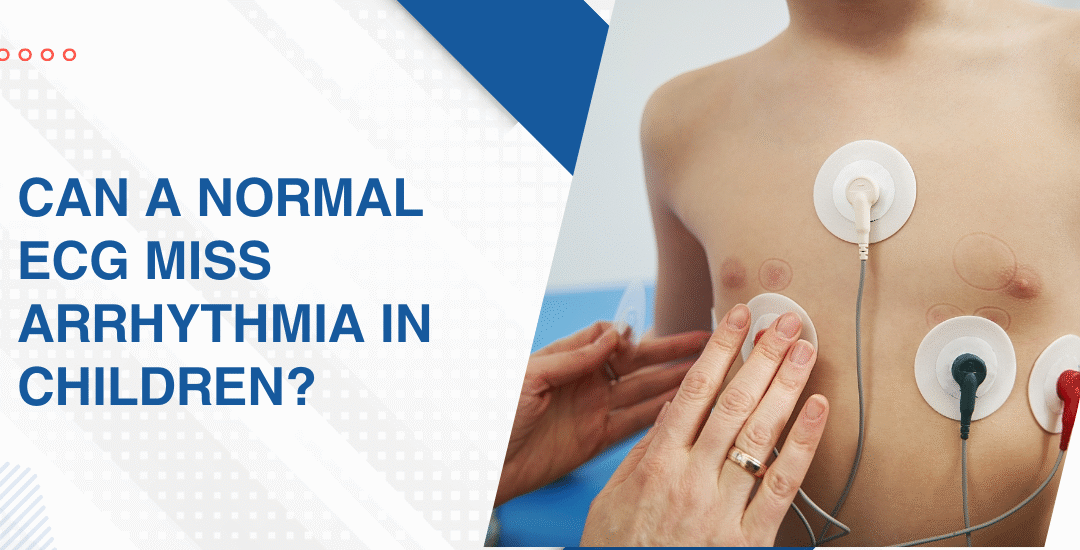
Can a Normal ECG Miss Arrhythmia in Children?
Yes, a normal resting ECG misses arrhythmias that are intermittent, exercise-triggered or only present during specific physiological states because it captures seconds of electrical activity from a heart that may only misbehave for minutes a day. A normal ECG tells you the rhythm was normal when the stickers were on. It says nothing about what happens the rest of the time.
“Parents bring in a normal ECG and think that’s the end of the investigation and I understand why but a child who faints during sport and has a normal resting ECG has not been cleared. They’ve had one test that was normal at rest. Those are completely different statements,” says Dr. Prashant Bobhate, Pediatric Cardiologist in Mumbai, India.
What Can a Normal Resting ECG Miss in Children?
More than most families realise. The ECG is twelve seconds long and arrhythmias don’t follow appointment schedules.
- Intermittent SVT: Supraventricular tachycardia episodes in children start and stop abruptly and can be completely absent at the time of an ECG meaning a child who had three episodes last week can have a perfectly normal tracing today and nobody would know from the paper alone.
- Long QT syndrome: Some long QT variants are borderline on a resting ECG especially in children whose QTc sits in the grey zone and the syndrome only declares itself as dangerous under adrenergic stress during exercise, sudden noise or emotional arousal that a resting trace never produces.
- Exercise-induced arrhythmia: Ventricular ectopics, sustained ventricular tachycardia and certain outflow tract arrhythmias only appear when heart rate rises with physical exertion and a resting ECG performed on a calm child lying still captures none of the electrical instability that exercise would immediately reveal.
- WPW with intermittent pre-excitation: In some children with Wolff-Parkinson-White syndrome the delta wave that indicates an accessory pathway is intermittent and can be absent on a resting ECG taken at a moment when normal conduction is dominant making the pathway invisible to a single snapshot investigation.
Every child with unexplained syncope, palpitations or exertional symptoms deserves more than a resting ECG and pediatric arrhythmia evaluation uses the right combination of investigations to catch what a single trace misses.
What Investigations Actually Catch Missed Arrhythmias in Children?
Once Tetralogy of Fallot is confirmed, the next steps focus on assessing severity, associated conditions, and planning the most appropriate treatment approach
- Severity determines urgency: A newborn with severe outflow obstruction and low saturations needs prostaglandin to keep the ductus open and urgent surgical planning while a baby with moderate obstruction and adequate saturations can be monitored and planned for elective repair in the first few months of life.
- Cardiac MRI in complex cases: When the pulmonary artery anatomy is complex or the echo leaves questions about branch pulmonary artery size or confluence a cardiac MRI or CT angiogram provides three-dimensional anatomy that guides the surgical approach in ways a two-dimensional echo can’t always match.
- Genetic testing: TOF is associated with 22q11 deletion syndrome and other chromosomal abnormalities often enough that genetic testing is recommended for every confirmed TOF newborn because the genetic result affects surgical risk, long term development planning and family counselling in ways that are missed entirely without testing.
- Surgical planning discussion: The timing, type of repair whether complete correction or a staged approach and the centre where surgery will be performed are all discussed with the family after the full diagnostic workup is complete and not before because the anatomy determines the plan not the diagnosis alone.
Parents wanting to understand what the earliest warning signs of critical cardiac disease look like in the newborn period before any formal diagnosis is made should read this piece on how to spot the early signs of heart disease in neonates because the earlier TOF is identified the more surgical options remain on the table and the less compromised the baby is when the operation happens.
Why Choose Dr. Prashant Bobhate for Paediatric Arrhythmia Assessment in Mumbai?
A child with unexplained syncope and a normal ECG needs a cardiologist who looks at the clinical history and immediately asks what the heart is doing when it isn’t in a clinic room and who knows which of the available investigations is most likely to capture the problem given how the symptoms present. Not a repeat resting ECG. The right next test. Dr. Prashant Bobhate has spent over 12 years managing paediatric arrhythmia across the full spectrum from benign ectopics through Long QT, WPW and complex ventricular arrhythmias requiring electrophysiology study at the Children’s Heart Centre, Kokilaben Dhirubhai Ambani Hospital. Escorts Heart Institute New Delhi.
Schedule a consultation to find out if a cure is possible and what the right treatment plan looks like for you.
FAQs
Can a child have a dangerous arrhythmia with a normal ECG?
Yes because conditions like Long QT syndrome, intermittent WPW and exercise-induced ventricular arrhythmias can all produce a completely normal resting ECG in between episodes that are genuinely dangerous when they occur.
What should parents do if their child faints but has a normal ECG?
Request further investigation because a normal ECG after syncope in a child is a starting point not a conclusion and Holter monitoring or an exercise stress test is usually the appropriate next step.
How long should a child wear a Holter monitor?
Usually 24 to 48 hours for daily or frequent symptoms but children with infrequent episodes need a longer event recorder worn for 30 days or more to capture the arrhythmia during an actual symptomatic episode.
Is a normal ECG enough before a child plays competitive sport?
Not always because exercise-induced arrhythmias and borderline Long QT syndrome require an exercise stress test for proper clearance and a resting ECG alone is insufficient screening for high-intensity competitive athletics in a symptomatic child.
References:
- Arrhythmia, MedlinePlus, U.S. National Library of Medicine — https://medlineplus.gov/arrhythmia.html
- Arrhythmia, National Heart Lung and Blood Institute — https://www.nhlbi.nih.gov/health/arrhythmias