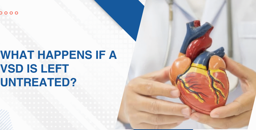
Is a Heart Murmur in a Child Dangerous?

Is a Heart Murmur in a Child Dangerous?

Most heart murmurs in children really aren’t the kind of thing worth panicking over, they tend to be innocent flow sounds the paediatrician catches during a routine check, and they usually fade on their own as the child grows. A smaller group, though, does hint at something structural underneath like a ventricular septal defect, a valve issue or a congenital shunt, and those are the ones a paediatric cardiologist needs to confirm or rule out on echo before anyone really starts to worry.

“Parents often panic the moment they hear the word murmur, but in most children it turns out to be a completely innocent finding, and even when it isn’t, catching it early gives us plenty of room to plan things calmly,” says Dr. Prashant Bobhate, Pediatric Cardiologist in Mumbai, India.

When Is a Heart Murmur in a Child Actually Dangerous?

Not every murmur carries clinical weight. But a handful of findings on examination do push one out of the reassurance bucket and straight into echo territory.

  • Loud, high-grade murmurs: Anything graded three or above on a six-point scale rarely ends up being innocent, because that kind of loudness usually means turbulent flow across something structural like a VSD or a tight valve, not the soft flow sound of a healthy heart.
  • Diastolic murmurs: A murmur heard between the two heart sounds is almost always pathological, since innocent ones only ever show up in systole when the heart contracts, so a diastolic finding in a child pushes the conversation straight toward structural assessment.
  • Murmurs sitting alongside worrying symptoms: Breathlessness during feeds, poor weight gain, a bluish tinge around the lips, repeated chest infections or a baby who tires out well before finishing a feed, any of these alongside a murmur point toward something real underneath.
  • Murmurs with genetic or family red flags: A family history of congenital heart disease, a known syndrome like Down or Turner, or a murmur that first turns up during a viral illness and hangs around well past the recovery, each is reason enough on its own to get a proper congenital heart disease workup done.

How Do Doctors Decide If a Child's Heart Murmur Needs Treatment?

The whole call really hinges on what the echo shows. A murmur by itself only tells you something inside the heart is making extra sound, not what’s causing it or whether the heart is coping with it.

  • Innocent murmur confirmed on echo: Needs nothing beyond reassurance and a note in the records, and most of these simply fade away by early adolescence without ever needing medication or another scan.
  • Small defects picked up on the side: A small VSD or a mild valve leak is usually watched rather than treated, with a repeat echo every six to twelve months, because many of these lesions either close on their own or stay small enough that they never really justify an intervention.
  • Moderate to large structural defects: These shift straight into active management, where the cardiologist weighs up catheter-based closure, medical therapy or surgical repair based on the lesion itself, the child’s age, and how well the heart is coping under load.
  • Murmurs with symptoms at any age: Any murmur turning up alongside breathlessness, poor growth, cyanosis or fatigue gets flagged as clinically important and worked up without delay, because symptomatic murmurs rarely turn out to be innocent once you look properly.

Parents wanting a deeper read on how structural defects actually show up in infancy, including the signs that often surface even before a murmur is picked up, can take a look at our piece on signs of a heart problem in a newborn baby, because catching the pattern early usually changes how much room there is to plan the next steps.

Why Choose Dr. Prashant Bobhate for Paediatric Heart Murmur Evaluation in Mumbai?

What really decides outcomes in a murmur case is the quality of the assessment itself, listening carefully, reading the echo against the full clinical picture, and then making a confident call on whether this is an innocent finding or something worth watching, instead of sending every family off for repeat imaging just to be safe. Dr. Prashant Bobhate has spent over 12 years evaluating paediatric heart murmurs across the full spectrum, from straightforward reassurance cases right through to complex congenital lesions, at the Children’s Heart Centre, Kokilaben Dhirubhai Ambani Hospital.

Schedule a consultation to understand what your child’s murmur actually means, and whether any further evaluation is genuinely needed from here.

FAQs

How do doctors tell an innocent murmur from a pathological one?

The clinician weighs up grade, timing, location and quality of the murmur, along with any associated symptoms or family history, and then confirms the impression on echocardiography whenever there’s any real doubt about whether the finding is truly innocent.

Do innocent heart murmurs in children need treatment?

Innocent murmurs need no medication, no restriction on activity, and no repeat imaging once an echo has shown the heart is structurally normal, and most of them just fade quietly by adolescence without ever coming back.

Can a heart murmur appear suddenly in a previously healthy child?

A new murmur can turn up during a fever or viral illness and disappear once the child recovers, though a persistent new murmur in an older child usually deserves a proper echo to rule out acquired valve involvement or rheumatic disease.

What tests are done to evaluate a child's heart murmur?

Evaluation typically starts with a detailed clinical examination, an ECG where it adds value, and 2D echocardiography as the main investigation, because echo gives a clear structural and functional picture of the heart in a completely non-invasive way.

References:

What Triggers Congenital Heart Defects?

What Triggers Congenital Heart Defects?

Congenital heart defects (CHDs) are primarily triggered by abnormal heart development within the first six weeks of pregnancy, often due to a combination of genetic factors, maternal health conditions, and environmental exposures. While many causes remain unknown, key factors include gene mutations, chromosome abnormalities, maternal infections, and substance use like smoking or alcohol.

“Most parents walk in convinced they did something wrong during the pregnancy, and honestly, in most cases the trigger was already written into the genes or tied to things nobody could have controlled,” says Dr. Prashant Bobhate, Pediatric Cardiologist in Mumbai, India.

What Are the Main Triggers of Congenital Heart Defects?

Triggers usually sit in three broad groups, genetic, maternal, and environmental, but in a real clinic they almost never turn up one at a time. More often, two or three of them are working on a heart that has very little wiggle room during those first delicate weeks of folding and chamber formation.

Chromosomal and genetic conditions tend to leave the clearest fingerprint of all. Down syndrome, DiGeorge, Turner, Noonan, each one brings its own cardiac signature, whether that shows up as an AV canal defect, a conotruncal anomaly or aortic coarctation, because the underlying genetic code throws off the careful sequence of cardiac looping inside the embryo.

Maternal diabetes is the next big piece of the puzzle, especially when sugars run high in the first trimester. Plenty of first-time mothers are caught off guard by this, but raised blood glucose during those early weeks quietly disturbs the molecular signals that guide the heart tube into its four-chambered shape.

Infections are the third thread to watch. Rubella in the first trimester is still the textbook example, and alongside it you’ve got CMV, influenza and a handful of other viral infections picked up while the foetal heart is still forming, each one capable of leaving behind damage that only really shows up once the baby is born.

The exposure side rounds it out, alcohol, retinoic acid, lithium, thalidomide, certain older anti-seizure drugs, any of these can disturb cardiogenesis if taken during the embryonic window. That’s exactly why something as ordinary as a preconception medication review matters a good deal more than most couples tend to realise before trying for a baby.

A child found to carry any of these triggers, whether on antenatal scan or after delivery, needs a proper congenital heart disease evaluation so the lesion is mapped out accurately and the long-term plan is shaped around what that one heart genuinely needs.

Why Do Some Babies Develop CHD Without Any Identifiable Trigger?

Quite a few CHD cases show up in pregnancies where nothing looks wrong on paper, and those are the hardest conversations, because parents come in hoping for a clean reason and medicine, being honest, cannot always give them one.

Most isolated defects come from multifactorial inheritance, a handful of low-effect genes nudging quietly against subtle environmental factors, none of which would cause harm alone, but together just enough to tip the heart past its threshold at a vulnerable moment. De novo mutations do something similar from a different angle, a fresh mutation neither parent carries appears in the embryo for the first time and throws cardiac development off course, which is exactly why a clean family history never reads as a true guarantee.

Then there’s the plain fact that cardiac morphogenesis is intricate work, precisely timed cell migrations and tissue fusions packed into a few short weeks, so small stochastic shifts in the process can quietly leave a defect behind even when every measurable factor looked completely fine. And every year, newer genetic and epigenetic contributors keep being added into the picture, so the unknown-cause group is slowly shrinking, although it still accounts for a very real slice of paediatric heart diagnoses seen in clinic today.

Parents wanting to understand how the defect itself, whatever the trigger behind it, tends to announce itself in those first few days should read our piece on signs of a heart problem in a newborn baby, because picking up the clinical picture early genuinely changes which management options stay open.

Why Choose Dr. Prashant Bobhate for Congenital Heart Disease Care in Mumbai?

Knowing what triggered the defect is really only half the story. What shapes the outcome is the assessment that follows, reading the echo against the clinical picture, working out whether the lesion needs surgical repair, a catheter-based device or just watchful follow-up, and then building a plan that moves with the child as they grow, instead of something copy-pasted from a textbook. Dr. Prashant Bobhate has spent over 12 years managing the full range of congenital heart disease, from antenatal diagnosis through to complex post-operative care, at the Children’s Heart Centre, Kokilaben Dhirubhai Ambani Hospital.

Schedule a consultation to understand what your child’s diagnosis actually means, and what the right care pathway looks like from here.

FAQs

Can lifestyle changes during pregnancy prevent congenital heart defects?

Good preconception care goes a long way, covering diabetes control, folic acid supplementation, rubella vaccination before conception and staying off alcohol or teratogenic drugs, though no single step on its own can guarantee prevention in every case.

Are congenital heart defects always inherited from parents?

Inheritance really only explains a small slice of cases, with most defects coming from de novo mutations, multifactorial influences, or causes that stay unidentified even after a thorough family workup has been done.

Does maternal age affect the risk of CHD in the baby?

Older maternal age does lift the risk a little, mostly through its link with chromosomal disorders like Down syndrome, which on their own already carry a well-defined cardiac risk profile worth knowing about.

Can a heart defect be detected during pregnancy?

Detection is usually possible through fetal echocardiography, typically done between 18 and 24 weeks, and most major structural defects get picked up on this scan when it’s performed in experienced hands.

References:

What Causes Arrhythmia In Infants And Children

What Causes Arrhythmia In Infants And Children

Arrhythmia in infants and children is caused by structural heart defects, genetic ion channel disorders, heart muscle inflammation, electrolyte imbalances and in some cases no identifiable cause at all. Some are present from birth. Others develop after cardiac surgery, viral illness or as the child grows and a substrate that was always there finally declares itself under the right trigger.

“The cause matters enormously because the treatment for an arrhythmia from a repaired congenital heart is completely different to one from a channelopathy and getting that distinction right early is what stops families spending years on the wrong management,” says Dr. Prashant Bobhate, Pediatric Cardiologist in Mumbai, India.

What are the main causes of Arrhythmia in Children?

The cause varies significantly by age, cardiac history and whether the heart is structurally normal or not.

  • Congenital heart disease: Structural defects and post-surgical scar tissue create arrhythmia substrates in chambers that were never built to manage the pressures and volumes they ended up carrying across years of abnormal haemodynamics.
  • Channelopathies: Long QT syndrome, Brugada syndrome and CPVT are genetic ion channel disorders that leave the heart structurally normal on echo but electrically unstable enough to trigger ventricular fibrillation under specific physiological conditions.
  • Myocarditis: Viral inflammation of the heart muscle disrupts normal conduction and creates zones of electrical instability that persist for months after the child looks and feels completely well again.
  • Post-surgical substrate: Scar tissue from atrial and ventricular incisions, patches and suture lines acts as the anatomical substrate for macro re-entrant arrhythmias including incisional atrial flutter that can develop years after a technically successful repair.

Every child with a documented or suspected arrhythmia needs a systematic assessment and pediatric arrhythmia evaluation maps the structural, electrical and genetic picture before any management decision is made.

What Causes Arrhythmia Specifically in Newborns and Young Infants?

Newborn arrhythmias have distinct causes that differ substantially from those seen in older children and each carries its own urgency and treatment path.

  • Accessory pathways: WPW and other accessory pathway tachycardias are among the most common causes of SVT in infants and the very fast rates they generate can cause heart failure within hours in a baby whose parents had no idea anything was wrong.
  • Congenital heart block: Maternal anti-Ro and anti-La antibodies cross the placenta and damage the fetal AV node causing complete heart block that needs pacemaker implantation in symptomatic neonates who can’t sustain adequate cardiac output at the slow ventricular rate.
  • Metabolic causes: Hypoglycaemia, hypocalcaemia and hyperkalaemia in sick neonates alter the electrical environment of the heart enough to produce rhythm disturbances that resolve completely once the metabolic abnormality is corrected without any antiarrhythmic treatment.
  • Idiopathic neonatal SVT: Many infants develop SVT in the first months of life with no identifiable structural genetic or metabolic cause and while these often resolve by twelve months the initial presentation can be dramatic enough to need emergency cardioversion.

Parents wanting to understand how cardiac abnormalities including arrhythmia substrates are detected in the newborn period before any symptomatic episode occurs should read this piece on newborn pulse oximetry heart screening because early detection changes what management options are available before a rhythm problem declares itself with a frightening episode.

Why Choose Dr. Prashant Bobhate for Paediatric Arrhythmia Care in Mumbai?

Finding the cause of arrhythmia in a child means reading the ECG in context, interpreting the Holter against the clinical history, deciding whether the echo is enough or whether genetic testing changes the management and putting all of that together into a plan that matches what this specific child’s rhythm problem actually requires. Not a protocol. A real assessment built around that one child. Dr. Prashant Bobhate has spent over 12 years managing paediatric arrhythmia across its full causal spectrum from structural post-surgical substrates through channelopathies through neonatal accessory pathway tachycardias at the Children’s Heart Centre, Kokilaben Dhirubhai Ambani Hospital.

Schedule a consultation to find out if a cure is possible and what the right treatment plan looks like for you.

FAQs

Can arrhythmia in infants resolve on its own?

Some can especially idiopathic neonatal SVT which often resolves by twelve months but channelopathies and post-surgical arrhythmias need ongoing management rather than watchful waiting.

Is arrhythmia in a newborn an emergency?

It depends on the rate and haemodynamic impact because a newborn in SVT at 280 beats per minute with poor perfusion is an emergency while a baby with isolated ectopic beats and normal function is not.


Does a child with arrhythmia need lifelong medication?

Not always because some arrhythmias are age-limited, some respond to ablation and some need only activity restriction but the decision depends entirely on the cause and the specific rhythm disorder identified.

Can genetic testing identify the cause of arrhythmia in children?

Yes for channelopathies including Long QT, Brugada and CPVT where genetic testing identifies the causative mutation and allows family members to be screened before they have a symptomatic episode.


References:

Can a Normal ECG Miss Arrhythmia in Children?

Can a Normal ECG Miss Arrhythmia in Children?

Yes, a  normal resting ECG misses arrhythmias that are intermittent, exercise-triggered or only present during specific physiological states because it captures seconds of electrical activity from a heart that may only misbehave for minutes a day. A normal ECG tells you the rhythm was normal when the stickers were on. It says nothing about what happens the rest of the time.

“Parents bring in a normal ECG and think that’s the end of the investigation and I understand why but a child who faints during sport and has a normal resting ECG has not been cleared. They’ve had one test that was normal at rest. Those are completely different statements,” says Dr. Prashant Bobhate, Pediatric Cardiologist in Mumbai, India.

What Can a Normal Resting ECG Miss in Children?

More than most families realise. The ECG is twelve seconds long and arrhythmias don’t follow appointment schedules.

  • Intermittent SVT: Supraventricular tachycardia episodes in children start and stop abruptly and can be completely absent at the time of an ECG meaning a child who had three episodes last week can have a perfectly normal tracing today and nobody would know from the paper alone.
  • Long QT syndrome: Some long QT variants are borderline on a resting ECG especially in children whose QTc sits in the grey zone and the syndrome only declares itself as dangerous under adrenergic stress during exercise, sudden noise or emotional arousal that a resting trace never produces.
  • Exercise-induced arrhythmia: Ventricular ectopics, sustained ventricular tachycardia and certain outflow tract arrhythmias only appear when heart rate rises with physical exertion and a resting ECG performed on a calm child lying still captures none of the electrical instability that exercise would immediately reveal.
  • WPW with intermittent pre-excitation: In some children with Wolff-Parkinson-White syndrome the delta wave that indicates an accessory pathway is intermittent and can be absent on a resting ECG taken at a moment when normal conduction is dominant making the pathway invisible to a single snapshot investigation.

Every child with unexplained syncope, palpitations or exertional symptoms deserves more than a resting ECG and pediatric arrhythmia evaluation uses the right combination of investigations to catch what a single trace misses.

What Investigations Actually Catch Missed Arrhythmias in Children?

Once Tetralogy of Fallot is confirmed, the next steps focus on assessing severity, associated conditions, and planning the most appropriate treatment approach

  • Severity determines urgency: A newborn with severe outflow obstruction and low saturations needs prostaglandin to keep the ductus open and urgent surgical planning while a baby with moderate obstruction and adequate saturations can be monitored and planned for elective repair in the first few months of life.
  • Cardiac MRI in complex cases: When the pulmonary artery anatomy is complex or the echo leaves questions about branch pulmonary artery size or confluence a cardiac MRI or CT angiogram provides three-dimensional anatomy that guides the surgical approach in ways a two-dimensional echo can’t always match.
  • Genetic testing: TOF is associated with 22q11 deletion syndrome and other chromosomal abnormalities often enough that genetic testing is recommended for every confirmed TOF newborn because the genetic result affects surgical risk, long term development planning and family counselling in ways that are missed entirely without testing.
  • Surgical planning discussion: The timing, type of repair whether complete correction or a staged approach and the centre where surgery will be performed are all discussed with the family after the full diagnostic workup is complete and not before because the anatomy determines the plan not the diagnosis alone.

Parents wanting to understand what the earliest warning signs of critical cardiac disease look like in the newborn period before any formal diagnosis is made should read this piece on how to spot the early signs of heart disease in neonates because the earlier TOF is identified the more surgical options remain on the table and the less compromised the baby is when the operation happens.

Why Choose Dr. Prashant Bobhate for Paediatric Arrhythmia Assessment in Mumbai?

A child with unexplained syncope and a normal ECG needs a cardiologist who looks at the clinical history and immediately asks what the heart is doing when it isn’t in a clinic room and who knows which of the available investigations is most likely to capture the problem given how the symptoms present. Not a repeat resting ECG. The right next test. Dr. Prashant Bobhate has spent over 12 years managing paediatric arrhythmia across the full spectrum from benign ectopics through Long QT, WPW and complex ventricular arrhythmias requiring electrophysiology study at the Children’s Heart Centre, Kokilaben Dhirubhai Ambani Hospital. Escorts Heart Institute New Delhi.

Schedule a consultation to find out if a cure is possible and what the right treatment plan looks like for you.

FAQs

Can a child have a dangerous arrhythmia with a normal ECG?

Yes because conditions like Long QT syndrome, intermittent WPW and exercise-induced ventricular arrhythmias can all produce a completely normal resting ECG in between episodes that are genuinely dangerous when they occur.

What should parents do if their child faints but has a normal ECG?

Request further investigation because a normal ECG after syncope in a child is a starting point not a conclusion and Holter monitoring or an exercise stress test is usually the appropriate next step.


How long should a child wear a Holter monitor?

Usually 24 to 48 hours for daily or frequent symptoms but children with infrequent episodes need a longer event recorder worn for 30 days or more to capture the arrhythmia during an actual symptomatic episode.

Is a normal ECG enough before a child plays competitive sport?

Not always because exercise-induced arrhythmias and borderline Long QT syndrome require an exercise stress test for proper clearance and a resting ECG alone is insufficient screening for high-intensity competitive athletics in a symptomatic child.


References:

Tests to Confirm Tetralogy of Fallot in a Newborn

Tests to Confirm Tetralogy of Fallot in a Newborn

Tetralogy of Fallot in a newborn is confirmed primarily through echocardiography which maps all four components of the defect including the VSD, right ventricular outflow tract obstruction, overriding aorta and right ventricular hypertrophy in detail. Supporting investigations include ECG, chest X-ray and pulse oximetry and together these give the cardiac team everything needed to plan surgical timing before the baby deteriorates.

“TOF in a newborn doesn’t always present dramatically. Some babies are pink enough to pass a routine examination and the diagnosis only comes when someone does an echo because the saturations were borderline or the murmur was unusual. That’s exactly why pulse oximetry screening in every newborn matters so much,” says Dr. Prashant Bobhate, Pediatric Cardiologist in Mumbai, India.

What Tests Confirm TOF in a Newborn and What Does Each Show?

Each investigation adds a different layer and together they build the full picture the surgical team needs before any planning conversation happens.

  • Echocardiography: The definitive test that maps the VSD size and location, measures the degree of right ventricular outflow tract obstruction, assesses pulmonary valve and artery anatomy and confirms the aortic override in enough detail to plan the surgical approach entirely from echo alone in most cases.
  • Chest X-ray: The classic boot-shaped cardiac silhouette from right ventricular enlargement and a concave pulmonary artery segment is often visible and reduced pulmonary vascular markings indicate reduced pulmonary blood flow that tells the team how obstructed the outflow actually is before the echo even begins.
  • ECG: Shows right ventricular hypertrophy with right axis deviation in most TOF cases and while it doesn’t confirm the anatomy it adds supporting electrical evidence and helps the team assess rhythm and conduction before planning anaesthesia and surgery.
  • Pulse oximetry: A pre and post-ductal saturation screen flags the oxygen desaturation pattern that TOF produces and in newborns where cyanosis isn’t yet visually obvious pulse oximetry is often the first investigation that pushes the neonatal team toward urgent cardiology referral rather than watchful observation.

Every newborn with a suspected or confirmed TOF diagnosis needs a detailed structural assessment and tetralogy of fallot evaluation maps the anatomy, the haemodynamic severity and the surgical timeline before any management decision is made.

What Happens After TOF Is Confirmed in a Newborn?

Once Tetralogy of Fallot is confirmed, the next steps focus on assessing severity, associated conditions, and planning the most appropriate treatment approach

  • Severity determines urgency: A newborn with severe outflow obstruction and low saturations needs prostaglandin to keep the ductus open and urgent surgical planning while a baby with moderate obstruction and adequate saturations can be monitored and planned for elective repair in the first few months of life.
  • Cardiac MRI in complex cases: When the pulmonary artery anatomy is complex or the echo leaves questions about branch pulmonary artery size or confluence a cardiac MRI or CT angiogram provides three-dimensional anatomy that guides the surgical approach in ways a two-dimensional echo can’t always match.
  • Genetic testing: TOF is associated with 22q11 deletion syndrome and other chromosomal abnormalities often enough that genetic testing is recommended for every confirmed TOF newborn because the genetic result affects surgical risk, long term development planning and family counselling in ways that are missed entirely without testing.
  • Surgical planning discussion: The timing, type of repair whether complete correction or a staged approach and the centre where surgery will be performed are all discussed with the family after the full diagnostic workup is complete and not before because the anatomy determines the plan not the diagnosis alone.

Parents wanting to understand what the earliest warning signs of critical cardiac disease look like in the newborn period before any formal diagnosis is made should read this piece on how to spot the early signs of heart disease in neonates because the earlier TOF is identified the more surgical options remain on the table and the less compromised the baby is when the operation happens.

Why Choose Dr. Prashant Bobhate for Cardiac Travel Clearance in Mumbai?

A pre-travel cardiac assessment for a child with CHD isn’t a rubber stamp. It’s a look at the current echo, the current saturations, the specific destination, the flight duration and the medical facilities available at the other end and then an honest answer about whether this trip is safe as planned or needs modification. Dr. Prashant Bobhate has spent over 12 years advising families with paediatric cardiac conditions on travel safety, pre-travel oxygen assessment and emergency planning across the full spectrum of congenital and pulmonary vascular disease at the Children’s Heart Centre, Kokilaben Dhirubhai Ambani Hospital.

Schedule a consultation to find out if a cure is possible and what the right treatment plan looks like for you.

FAQs

Is echocardiography always needed to confirm TOF in a newborn?

Yes, because clinical examination and chest X-ray can raise suspicion but only echo maps the four components of TOF in enough detail to confirm the diagnosis and plan surgery.

Can TOF be missed on a routine newborn examination?

Yes especially in pink TOF where cyanosis isn’t visible and no murmur is audible yet which is exactly why pulse oximetry screening in every newborn is the safety net that catches these cases before discharge.


Does every newborn with TOF need immediate surgery?

No because timing depends on the degree of outflow obstruction and oxygen saturations and some babies are stabilised and planned for elective repair in the first few months while others need urgent intervention from the first days of life.

Does a PDA murmur always mean the defect is significant?

No because murmur intensity doesn’t reliably correlate with shunt size in premature babies and a soft or absent murmur can accompany a large haemodynamically significant PDA which is exactly why echo is always needed rather than clinical assessment alone.


References:

Call Now Button