What Causes Sudden Cardiac Death In Young Athletes?
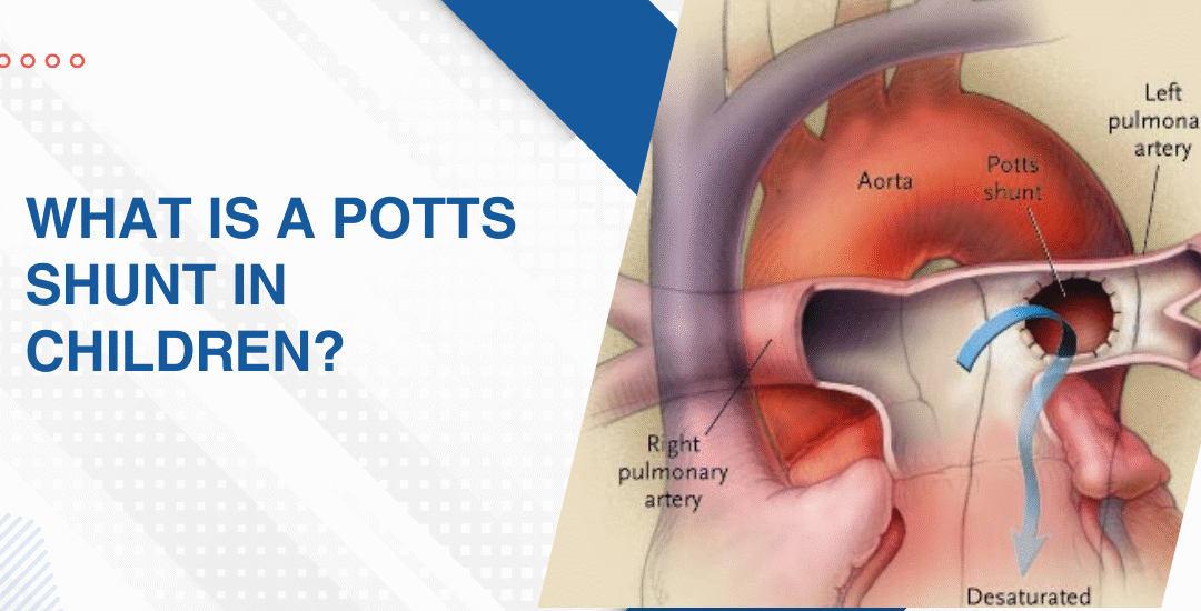
A diet for children with pulmonary hypertension should focus on low-sodium, nutrient-dense whole foods that support heart and lung function. Key priorities include limiting processed food and added salt, ensuring adequate protein for muscle strength, managing total fluid intake and maximising vitamins C, D and iron.
“Families focus almost entirely on medications and rightly so but nutrition is the one thing happening three times a day every day that nobody is optimising and in a child with pulmonary hypertension that’s a significant missed opportunity for supporting the heart,” says Dr. Prashant Bobhate, Pediatric Cardiologist in Mumbai, India.
What Are the Most Common Causes of Sudden Cardiac Death in Young Athletes?
There’s a recognisable list and understanding it is what makes pre-participation cardiac screening a clinical necessity rather than an optional extra for competitive sport.
- Hypertrophic cardiomyopathy: The single most common cause in young athletes where abnormally thickened myocardial fibres create an unstable electrical substrate that generates ventricular fibrillation under the precise haemodynamic stress that intense physical exertion produces.
- Anomalous coronary arteries: A coronary artery arising from the wrong sinus can course between the aorta and pulmonary artery in a way that compresses the vessel during exercise-induced aortic expansion and cuts off blood supply to a large myocardial territory at exactly the moment demand is highest.
- Long QT syndrome: The ECG looks almost normal at rest but the prolonged repolarisation interval becomes dangerous under adrenergic stimulation from exercise or sudden emotional stress and can trigger torsades de pointes that degenerates into ventricular fibrillation without any warning symptom.
- Myocarditis: Viral inflammation of the heart muscle creates zones of electrical instability that persist well beyond the acute illness phase and an athlete who returns to training too soon after a viral illness with unrecognised myocarditis is training on a heart that can no longer reliably maintain normal rhythm under load.
Every young athlete with a family history of sudden cardiac death, unexplained syncope during exercise or a newly detected murmur deserves a formal assessment and pediatric arrhythmia evaluation maps the electrical and structural risk before any return to competitive sport gets cleared.
Can Sudden Cardiac Death in Young Athletes Be Prevented?
Often yes. When the right screening happens before the right sport at the right intensity.
- Pre-participation ECG: A resting 12-lead ECG picks up Long QT syndrome, WPW pattern, Brugada pattern and HCM-related repolarisation changes that a physical examination alone completely misses and costs a fraction of what treating a survivor of cardiac arrest costs in every dimension imaginable.
- Echocardiography for high-risk athletes: Any athlete with an abnormal ECG, a family history of sudden cardiac death under 50, unexplained exertional syncope or a cardiac murmur needs an echo before competing at any level where sustained high-intensity effort is involved.
- Restricting sport in diagnosed conditions: A child diagnosed with HCM, Long QT or anomalous coronary arteries needs a formal sport eligibility assessment because the specific activity restriction required is condition-specific and a blanket ban applied without assessment is as clinically unhelpful as no restriction at all.
- AED availability at sports venues: Automated external defibrillators at schools, sports academies and training grounds don’t prevent the arrhythmia but they convert a potentially fatal event into a survivable one when used within the first three to five minutes and their absence at Indian sports venues is a preventable gap that costs young lives every year.
Parents wanting to understand what cardiac warning signs in young children look like before any sport-related event forces the issue should read this piece on how to spot the early signs of heart disease in neonates because the cardiac conditions that cause sudden death in athletes at sixteen were present at birth and detectable long before the first sprint.
Why Choose Dr. Prashant Bobhate for PH Management in Mumbai?
Pre-participation cardiac screening for a young athlete isn’t a routine check. It’s reading an ECG in the context of the sport being played, the intensity involved and the specific conditions that that particular child’s family history or symptom profile makes worth looking for. Not a form-filling exercise. A real assessment. Dr. Prashant Bobhate has spent over 12 years managing paediatric arrhythmia, hypertrophic cardiomyopathy, sudden cardiac death risk stratification and sport eligibility assessments across every age group at the Children’s Heart Centre, Kokilaben Dhirubhai Ambani Hospital. Escorts Heart Institute New Delhi.
Schedule a consultation to find out if a cure is possible and what the right treatment plan looks like for you.
FAQs
How common is sudden cardiac death in young athletes in India?
Exact figures are underreported because many events are attributed to heat stroke or exhaustion without cardiac investigation but HCM and arrhythmia syndromes are consistently identified as the leading causes wherever proper post-event evaluation is carried out.
Should every child have a cardiac screening before playing competitive sport?
Yes at minimum an ECG and clinical history because the conditions that cause sudden cardiac death in athletes are detectable before the event and a normal screening is a genuinely reassuring baseline that parents and coaches both deserve to have.
Can a child with a heart condition ever play sport?
Many can with the right assessment and specific guidance because not every cardiac condition carries the same exercise risk and a formal sport eligibility assessment gives a child and family a real answer rather than a blanket restriction based on diagnosis alone.
What should parents do if their child faints during sport?
Treat it as a cardiac event until proven otherwise because exertional syncope in a young athlete is always a red flag that needs same day cardiac assessment including ECG and echo before the child returns to any physical activity.
References:
- Arrhythmia, MedlinePlus, U.S. National Library of Medicine — https://medlineplus.gov/arrhythmia.html
- Hypertrophic Cardiomyopathy, National Heart Lung and Blood Institute — https://www.nhlbi.nih.gov/health/cardiomyopathy