What Is Tetralogy of Fallot?

What Is Tetralogy of Fallot?

Tetralogy of Fallot is a congenital heart condition made up of four specific structural defects that occur together inside the heart from birth. It reduces the oxygen reaching the blood and it’s one of the most common serious heart defects found in children. Scary name. But genuinely one of the most treatable cardiac diagnoses a child can receive.

“Tetralogy of Fallot sounds overwhelming the first time you hear it but these four defects are well understood and children who get the right surgical repair at the right time do genuinely well,” says Dr. Prashant Bobhate, Pediatric Cardiologist in Mumbai, India.

What Are the Four Defects That Make Up Tetralogy of Fallot?

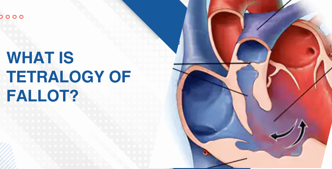
The name literally means four. And understanding what each of those four things does to blood flow inside your child’s heart is what makes everything a specialist says from that point forward actually land properly.

  • Ventricular septal defect: A hole between the two lower chambers lets oxygen-poor blood mix with oxygen-rich blood before it even leaves the heart and that mixing is exactly what drives the low oxygen levels that show up on the skin and lips as that bluish colour most families notice first and can’t unsee after.
  • Pulmonary stenosis: The valve leading from the heart toward the lungs is narrowed and sometimes the entire passage below it is narrowed too which means the right side of the heart is pushing far harder than it was ever built to just to get blood through to the lungs at all.
  • Right ventricular hypertrophy: Because the right side works so much harder against that narrowed outflow the muscle wall thickens trying to keep up. It’s the heart doing its absolute best to compensate for a situation it genuinely cannot fix on its own no matter how hard it tries.
  • Overriding aorta: The aorta sits shifted directly over the hole between the chambers instead of sitting entirely over the left side where it belongs and this means it pulls blood from both chambers simultaneously rather than only the clean oxygenated blood it was designed to carry.

These four things together are what makes Tetralogy of Fallot a condition that needs surgical correction rather than watchful waiting and the earlier that correction happens the better and faster the heart bounces back from it.

What Are the Signs of Tetralogy of Fallot and How Is It Treated?

Some children with TOF are diagnosed before birth. Others come to attention in those first fragile weeks at home. And occasionally a milder form gets missed entirely until a child is older and something just keeps not adding up no matter how many times it gets explained away.

  • The blue spells nobody forgets: Tet spells are sudden episodes where the baby turns deeply blue, becomes distressed or goes floppy and they happen when the narrowed pulmonary outflow drops blood oxygen dramatically in a way that doesn’t wait for a convenient moment to do it.
  • Squatting in older children: A child with unrepaired TOF who instinctively squats during activity is doing something physiologically clever without knowing it because squatting increases lower body resistance and briefly pushes more blood toward the lungs to boost oxygen when the body needs it most.
  • Surgical repair usually between 3 and 6 months: Most centres repair TOF in the first six months of life with a complete repair that closes the VSD, widens the pulmonary outflow and repositions the aorta all in one operation rather than staging it across multiple procedures over years.
  • Outcomes after repair are excellent: Children who get complete TOF repair at the right time grow up to live normal active lives and while lifelong cardiac follow up is part of the picture the vast majority reach adulthood with genuinely good heart function and no meaningful limitations on how they live.

Parents wanting to understand what early cardiac warning signs actually look like before any diagnosis gets made should read this piece on how to spot the early signs of heart disease in neonates which goes through what these signs genuinely look like in those first weeks at home when everything still feels new and impossible to interpret properly.

Why Choose Dr. Prashant Bobhate for Tetralogy of Fallot Care in Mumbai?

A TOF diagnosis needs someone who has spent years specifically inside complex congenital cardiac disease. Not someone applying a general protocol to a condition that has never once responded well to general anything. Dr. Prashant Bobhate trained at Escorts Heart Institute in New Delhi then went deliberately to the University of Alberta in Canada for advanced paediatric cardiac fellowship training. Over 12 years working with children at every stage and complexity of congenital heart disease. His team performed India’s very first successful Transcatheter Potts Shunt and actively manages over 400 children on advanced therapy right now. He doesn’t give families a standard answer pulled from a protocol sheet. He looks at your child’s specific anatomy and builds the plan entirely from what he actually finds there.

Schedule a consultation to find out if a cure is possible and what the right treatment plan looks like for you.

FAQs

Can TOF be detected before birth?

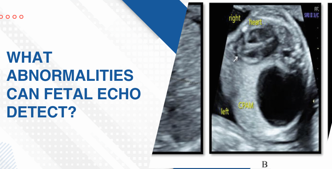
Yes. Fetal echocardiography between 18 and 24 weeks can identify TOF before birth which allows families to plan delivery at a specialist centre with a paediatric cardiac team ready from the very first moment rather than scrambling after the fact.

Is surgery always needed for TOF?

Yes always. TOF can’t be managed with medication alone and complete surgical repair closing the VSD and widening the pulmonary outflow is the only thing that actually resolves the four underlying structural problems properly.

Can a child with repaired TOF play sports?

Most can participate in normal physical activity and the specific level of exercise allowed is determined through regular cardiac follow up rather than a blanket restriction applied to every child the same way regardless of their individual situation.

What's the long term outlook after TOF repair?

Excellent for the vast majority. Most children who get complete repair in infancy grow into adults with good heart function though lifelong follow up is needed to monitor the pulmonary valve and right heart as they get older.

References:

Is Fetal Echocardiogram Safe for the Baby

Is Fetal Echocardiogram Safe for the Baby

Yes. A fetal echocardiogram is completely safe for both the baby and the mother. It uses standard ultrasound technology with no radiation involved and no known risk at any stage of pregnancy. The same technology that’s been used in routine pregnancy scans for decades. Just applied more carefully and for longer to look specifically at the heart.

“A fetal echo uses the exact same ultrasound waves as a routine pregnancy scan. There is no radiation. No risk to the baby. Just a longer, more detailed look at a heart that deserves that attention,” says Dr. Prashant Bobhate, Pediatric Cardiologist in Mumbai, India.

Why Is a Fetal Echo Safe and What Does It Actually Involve?

Most parents asking this question aren’t really asking about physics. They’re asking whether something is going to hurt their baby. And the honest answer is no. Nothing about this scan puts your baby at risk in any way.

  • No radiation whatsoever: Unlike X-rays or CT scans a fetal echocardiogram uses only sound waves to build its images and sound waves don’t damage tissue, don’t affect developing cells and don’t accumulate in the body in a way that causes any problem at all.
  • Same technology as a routine scan: The transducer pressed against the abdomen is identical to what gets used in every standard pregnancy ultrasound and the only difference is the operator’s focus, the duration and how carefully those heart structures get examined frame by frame.
  • Can be repeated without concern: If the baby’s position makes imaging difficult the first time or if a follow up scan is needed to monitor a finding there’s no limit on how many times a fetal echo can be safely done and no cumulative risk that builds up with repeat scans.
  • No needles, no sedation, nothing invasive: The entire scan happens externally through the mother’s abdomen and in some cases a transvaginal probe is used for earlier scans but nothing enters the uterus, nothing touches the baby and nothing about the process is invasive at any point.

Understanding what the scan actually involves is what makes fetal echocardiography feel like something you’re walking into with your eyes open rather than something that was just added to your appointment list without any real explanation.

What Should You Actually Expect During and After a Fetal Echo?

Because the safety question is usually followed by a practical one. What actually happens in that room. How long is it. Will something feel wrong. And what do you do if the scan finds something you weren’t expecting to hear about that day.

  • The scan takes 30 to 60 minutes: Longer than a routine anomaly scan because the cardiologist is examining every chamber, every valve, every vessel and watching blood flow in real time rather than taking a quick look and moving on to the next item on the checklist.
  • You’ll feel nothing unusual: Warm gel on the abdomen, a transducer pressed gently against the skin and a lot of quiet concentration from the person doing the scan. Some babies cooperate immediately. Others take their time settling into a position that shows what needs to be seen.
  • Results are usually discussed the same day: A paediatric cardiologist performing the scan can give you a clear picture of what was found or what wasn’t found before you leave the room and that conversation is often the part families feel most unprepared for regardless of what the answer turns out to be.
  • A normal result is genuinely reassuring: Not a perfect guarantee because some very small defects only become visible after birth but a normal fetal echo from an experienced specialist means the vast majority of significant structural problems have been looked for and not found.

Parents in a high risk category wanting to understand what happens if something does get found during the scan should read this piece on importance of fetal diagnosis of critical congenital heart disease which explains honestly what an early prenatal finding actually changes about the care plan and the options available to families from that point.

Why Choose Dr. Prashant Bobhate for Fetal Echocardiography in Mumbai?

A fetal echo being safe doesn’t mean every fetal echo is equally useful. The safety is in the technology. The value is entirely in the person reading it. Dr. Prashant Bobhate trained at Escorts Heart Institute in New Delhi then went specifically to the University of Alberta in Canada for advanced paediatric cardiac fellowship training. Over 12 years reading these scans and then following those same babies through every stage of what came after. His team performed India’s very first successful Transcatheter Potts Shunt and actively manages over 400 children on advanced therapy right now. He doesn’t just tell you the scan was normal or abnormal. He tells you what it actually means for your specific baby and what needs to happen next because of it.

Schedule a consultation to find out if a cure is possible and what the right treatment plan looks like for you.

FAQs

Does fetal echo use radiation?

No. It uses only sound waves exactly like a routine pregnancy ultrasound and there is no radiation involved at any point which means there is no radiation related risk to the baby or mother whatsoever.

Can fetal echo be done multiple times in one pregnancy?

Yes without any concern. There is no cumulative risk from repeat fetal echocardiograms and if follow up scans are needed to monitor a finding they can be scheduled as many times as clinically necessary.

Does the baby feel anything during the scan?

No. The scan is entirely external through the mother’s abdomen and uses sound waves that the baby cannot feel in any way that differs from the ordinary sound environment of the womb.

What if my baby moves too much during the scan?

The sonographer waits for a better position and the scan continues when the baby settles because image quality matters more than speed and no part of the scan can be rushed without compromising what can actually be seen.

References:

Is Congenital Heart Disease Hereditary

Is Congenital Heart Disease Hereditary

Congenital heart disease can be hereditary but it isn’t always. Some types carry a clear genetic link and run through families for generations. Others show up with zero family history anywhere. Most cases are actually a mix of genetic vulnerability and something that happened in those first few weeks of pregnancy before most people even knew they were pregnant.

“Genetics plays a role in congenital heart disease but it’s rarely the whole story. Understanding your specific risk is what allows families to plan rather than just worry,” says Dr. Prashant Bobhate, Pediatric Cardiologist in Mumbai, India.

What Is the Genetic and Hereditary Link in Congenital Heart Disease?

This question doesn’t stay academic for long. The moment a cardiac diagnosis lands in your family it becomes the most personal thing in the room.

  • Single gene mutations: Noonan syndrome, Marfan syndrome, DiGeorge syndrome. Each one carries a specific mutation that raises structural heart defect risk significantly and each one can pass from parent to child in a pattern a genetic counsellor can actually trace through your specific family rather than just quoting you a percentage and sending you home.
  • Chromosomal abnormalities: Down syndrome, Turner syndrome, trisomy 18. All of them carry substantially elevated rates of congenital heart findings and in these cases the cardiac piece isn’t standalone at all it’s part of a much broader picture that genuinely needs to be understood as a whole thing not in fragments.
  • Familial recurrence risk: If one parent has congenital heart disease their child’s risk sits around 3 to 5 percent. Compare that to under 1 percent in the general population. And if a previous child was already affected that number shifts again depending entirely on which specific defect we’re actually talking about.
  • Multifactorial inheritance: Here’s the honest part most families don’t hear clearly enough. Most congenital heart cases don’t follow a clean genetic pattern. They’re a combination of multiple small genetic variants mixing with environmental exposures during those first eight weeks when the heart is literally being built from nothing.

That specific picture in your family is what makes the right congenital heart disease planning conversation actually useful rather than just vaguely reassuring in a way that doesn’t change anything practically for you.

What Non-Genetic Factors Can Cause Congenital Heart Disease?

Because plenty of families with no cardiac history anywhere end up sitting in a paediatric cardiology clinic. 

  • Maternal diabetes in the first trimester: Poorly controlled blood sugar during those first twelve weeks more than triples the baseline risk of a structural heart defect forming and it happens during the exact window when the cardiac chambers and valves are being built completely from scratch for the very first time.
  • Certain medications taken early: Lithium, some antiepileptics, heavy alcohol during the first trimester. All of them carry documented associations with specific cardiac defect types and none of it has anything to do with what does or doesn’t run in your family.
  • Rubella and certain infections: This one still surprises people. A rubella infection in the first trimester remains one of the oldest known causes of congenital heart disease and it’s still relevant today wherever vaccination coverage isn’t as solid as the numbers on paper suggest it should be.
  • Maternal autoimmune conditions like lupus: Antibodies can cross the placenta and quietly interfere with the baby’s cardiac conduction system producing problems that look genetic on the surface but trace back to something completely different when you actually follow the thread properly.

Parents who received a congenital heart finding before birth and want to understand what early detection actually changes should read this piece on importance of fetal diagnosis of critical congenital heart disease which goes through honestly what a prenatal diagnosis means for the care plan and the options available from that point forward.

Why Choose Dr. Prashant Bobhate for Congenital Heart Disease Care in Mumbai?

Understanding whether what’s in your family carries a hereditary pattern isn’t something you want answered by someone who encounters this occasionally and figures it out as they go. Dr. Prashant Bobhate spent over 12 years specifically inside congenital cardiac disease in children. Fetal diagnosis. Intervention. Long term follow up. All of it. Trained at Escorts Heart Institute in New Delhi then went deliberately to the University of Alberta in Canada for advanced paediatric cardiac fellowship training. His team performed India’s very first successful Transcatheter Potts Shunt and actively manages over 400 children on advanced therapy right now. He doesn’t hand families a generic recurrence number from a textbook. He looks at your specific defect, your specific family history and builds the actual picture entirely from there.

Schedule a consultation to find out if a cure is possible and what the right treatment plan looks like for you.

FAQs

Next child at risk if first had CHD?

Recurrence risk depends entirely on the specific defect and whether a genetic cause has been found and a proper cardiac and genetic evaluation gives you a real number rather than a general guess that doesn’t actually help you plan anything.

Should I get genetic testing if CHD runs in my family?

Yes. Genetic counselling before or during a subsequent pregnancy helps identify whether a specific inheritable cause is present and whether fetal echocardiography between 18 and 24 weeks needs to be part of how that pregnancy gets monitored properly.

Can CHD skip a generation?

Some genetic forms can appear to skip generations entirely depending on how the specific mutation expresses itself across family members and that’s exactly why a full family cardiac history matters when you’re trying to understand what the real recurrence risk actually looks like.

Higher risk if both parents have heart conditions?

Yes and meaningfully so. When both parents carry cardiac history the recurrence risk is higher than when only one does and a specialist evaluation before the next pregnancy gives the clearest possible picture of what that combined risk means for your specific situation.

References:

What Abnormalities Can Fetal Echo Detect?

What Abnormalities Can Fetal Echo Detect?

A fetal echocardiogram can detect structural heart defects, abnormal blood flow patterns, valve problems, rhythm disturbances and major vessel abnormalities in an unborn baby. It gives a far more detailed picture of your baby’s heart than a routine anomaly scan ever will. And that difference in detail is exactly why it exists as a separate scan entirely.

“A fetal echo doesn’t just look at the heart. It reads how the heart is working, how blood is moving through it and whether anything is building that needs to be caught right now rather than after birth,” says Dr. Prashant Bobhate, Pediatric Cardiologist in Mumbai, India.

What Structural Heart Abnormalities Can a Fetal Echo Detect?

Most parents walking into this scan don’t know how much it can actually see. A routine 20-week anomaly scan checks the heart briefly. 

  • Ventricular septal defects: Holes between the lower chambers of the heart that let blood cross where it shouldn’t and while small ones sometimes close on their own larger ones need to be on the radar before birth so nothing about the delivery or immediate care comes as a surprise.
  • Transposition of the great arteries: The two main arteries leaving the heart have completely swapped positions and this is genuinely one of those findings where catching it before birth rather than in the first hours of life changes the entire outcome for that baby.
  • Hypoplastic left heart syndrome: The entire left side of the heart is severely underdeveloped and without a fetal diagnosis this one walks into the delivery room completely unannounced and the difference in survival between a prepared team and an unprepared one is stark.
  • Pulmonary and aortic valve abnormalities: Valves that are too narrow, leaking or structurally malformed in ways that affect how blood moves in and out of the heart and ones that a routine scan simply doesn’t have the resolution or the time to assess properly.

These are exactly the kinds of findings that make fetal echocardiography one of the most important scans a high risk pregnancy can have rather than an optional extra someone added to the list.

What Rhythm and Flow Abnormalities Can a Fetal Echo Pick Up?

Because it isn’t just about structure. A heart can be built correctly and still not work the way it should. And some of the most important things a fetal echo finds aren’t holes or missing pieces but problems with how the heart is actually beating and moving blood around.

  • Fetal arrhythmias: Heartbeats that are too fast, too slow or completely irregular in a pattern that no routine midwife check would ever catch properly and some of these need treatment that starts before birth rather than waiting until after delivery.
  • Abnormal blood flow across valves: Doppler imaging inside the fetal echo shows exactly how fast blood is moving and in which direction and flow that looks wrong is often the first sign of a valve problem that would otherwise stay invisible until symptoms appeared months later.
  • Fetal heart block: The electrical signals telling the heart to beat aren’t getting through properly and this can be connected to maternal autoimmune conditions the mother didn’t even know were affecting her baby’s cardiac conduction system in the background.
  • Tumours and structural masses: Cardiac rhabdomyomas sometimes spotted incidentally during a fetal echo and while most are benign they carry associations with other conditions that need to be on the clinical radar before birth rather than discovered afterwards entirely by accident.

Parents wanting to understand what happens when one of these findings does come up before birth should read this piece on importance of fetal diagnosis of critical congenital heart disease which goes through honestly what early detection actually changes about the care plan and the options available to families from that point forward.

Why Choose Dr. Prashant Bobhate for Fetal Echocardiography in Mumbai?

Finding something on a fetal echo is only half of it. Understanding what it means and what needs to happen next is where the real experience shows. Dr. Prashant Bobhate didn’t come to this area by accident. He trained at Escorts Heart Institute in New Delhi then went specifically to the University of Alberta in Canada for advanced paediatric cardiac fellowship training. Over 12 years reading these scans, following those babies through birth, through intervention and through recovery. His team performed India’s very first successful Transcatheter Potts Shunt and actively manages over 400 children on advanced therapy right now. He doesn’t just report what he sees. He tells you what it means for your specific baby.

Schedule a consultation to find out if a cure is possible and what the right treatment plan looks like for you.

FAQs

Can fetal echo detect all heart defects?

Most major structural defects can be detected but very small defects, minor valve abnormalities and certain muscular VSDs can sometimes only become visible after birth when the heart is larger and easier to image properly.

Is fetal echo safe for the baby?

Yes completely. It uses the same ultrasound technology as a routine pregnancy scan with no radiation involved and no known risk to the baby or mother at any gestational age it is performed.

What happens after an abnormality is found on fetal echo?

The family gets counselled about the specific finding, a delivery plan gets made at a specialist centre with the right neonatal cardiac team available and in some cases the treatment plan begins within hours of birth rather than days.

Does a normal fetal echo guarantee a healthy heart after birth?

A normal fetal echo is genuinely reassuring but it cannot guarantee every possible cardiac finding because some conditions develop or become detectable only after birth once the circulation has completed its normal postnatal transition.

References:

What Week of Pregnancy Is Fetal Echo Recommended?

What Week of Pregnancy Is Fetal Echo Recommended?

A fetal echocardiogram is generally recommended between 18 and 24 weeks of pregnancy. That’s when your baby’s heart is developed enough to show everything that needs to be seen clearly. Done too early and the picture is incomplete. Done too late and the imaging gets harder. The window is real and the timing genuinely matters.

“The timing of a fetal echocardiogram isn’t a suggestion. It’s the difference between getting a clear picture of your baby’s heart and getting an incomplete one that raises more questions than it answers,” says Dr. Prashant Bobhate, Pediatric Cardiologist in Mumbai, India

What Is the Ideal Week for a Fetal Echo and Why Does Timing Matter So Much?

Most parents assume any week is fine as long as the scan happens before birth. It isn’t. What a specialist can reliably see changes dramatically depending on exactly when you walk through that door.

  • 18 to 24 weeks is the gold standard: The heart is developed enough at this stage to show all four chambers clearly, the valves, the major vessels and the way blood is actually flowing through all of it in real time without guessing.
  • Earlier than 18 weeks misses things: A scan done at 14 or 15 weeks might catch the most obvious structural problems but smaller defects and valve abnormalities genuinely cannot be seen clearly enough to assess properly at that gestational age.
  • Later than 24 weeks has its own problems: The baby is larger, harder to position and the acoustic window through the mother’s abdomen gets more difficult which means image quality drops and what can be confirmed reliably starts shrinking.
  • 20 to 22 weeks hits the sweet spot: Most specialists will tell you this is the ideal range because the heart is fully formed enough to examine comprehensively while the baby is still small enough to get the imaging angles that actually show what needs to be seen.

Understanding exactly when your fetal echocardiography needs to happen is what makes the difference between a scan that genuinely answers your questions and one that leaves everyone uncertain about what was and wasn’t seen.

Who Actually Needs a Fetal Echo and When Should They Book It?

Not every pregnancy gets referred for this and a lot of parents who should be getting one never do because nobody flagged the risk factor sitting right there in their medical history.

  • Family history of heart defects: If you or your partner or a previous child had a congenital heart problem that risk is elevated enough that a fetal echo between 20 and 22 weeks isn’t optional it’s genuinely necessary.
  • Maternal diabetes or lupus: Both conditions affect fetal cardiac development in ways that don’t always show on a routine anomaly scan and both warrant a dedicated cardiac look at the right gestational window.
  • Abnormal findings on routine scan: An anomaly scan that flagged anything suspicious about the heart, the vessels or the rhythm is a direct referral indication and waiting to see if the next scan looks better is not the right response.
  • IVF pregnancies and multiple gestations: Both carry elevated rates of congenital heart findings compared to spontaneous singleton pregnancies and both deserve a fetal echo rather than relying on routine screening alone to catch what’s there.

Parents already in a high risk category wanting to understand what happens when a problem does get found before birth should read this piece on importance of fetal diagnosis of critical congenital heart disease which explains honestly what early detection actually changes about the options available to families.

Why Choose Dr. Prashant Bobhate for Fetal Echocardiography in Mumbai?

A fetal echo is only as useful as the person interpreting what’s on that screen and that’s not a small distinction when it’s your baby’s heart being assessed before birth. Dr. Prashant Bobhate trained at Escorts Heart Institute in New Delhi then went specifically to the University of Alberta in Canada for advanced paediatric cardiac fellowship training. Over 12 years working with babies from before birth through every stage and complexity of congenital heart disease.

Schedule a consultation to find out if a cure is possible and what the right treatment plan looks like for you.

FAQs

What is the best week for fetal echocardiography during pregnancy?

Between 20 and 22 weeks is the ideal window when the heart is fully developed enough to assess comprehensively and the baby is still positioned well enough to get clear imaging angles.

Can fetal echo be done earlier than 18 weeks of pregnancy?

An early fetal echo between 13 and 16 weeks is possible in very high risk cases but it cannot replace the standard 20 to 22 week scan because smaller defects simply cannot be reliably seen that early.

Does every pregnancy need a fetal echocardiogram?

Not every pregnancy but any pregnancy with elevated risk factors including family history of heart defects, maternal diabetes, abnormal anomaly scan findings or IVF conception should have one.

What happens if a heart defect is found on fetal echocardiography?

Finding a defect before birth gives families time to prepare and plan rather than facing a cardiac crisis in the delivery room.

References:

Call Now Button